An Integrated Textbook of Basic Science, Medicine, and Surgery
Hans G. Beger, Andrew L. Warshaw, Ralph H. Hruban, Markus W. Buchler, Markus M. Lerch, John P. Neoptolemos, Tooru Shimosegawa, David C. Whitcomb, Hans G. Beger, Andrew L. Warshaw, Ralph H. Hruban, Markus W. Buchler, Markus M. Lerch, John P. Neoptolemos, Tooru Shimosegawa, David C. Whitcomb
This is a test
This is a test
Condividi libro
English
ePUB (disponibile sull'app)
Disponibile su iOS e Android
eBook - ePub
The Pancreas
An Integrated Textbook of Basic Science, Medicine, and Surgery
Hans G. Beger, Andrew L. Warshaw, Ralph H. Hruban, Markus W. Buchler, Markus M. Lerch, John P. Neoptolemos, Tooru Shimosegawa, David C. Whitcomb, Hans G. Beger, Andrew L. Warshaw, Ralph H. Hruban, Markus W. Buchler, Markus M. Lerch, John P. Neoptolemos, Tooru Shimosegawa, David C. Whitcomb
Dettagli del libro
Anteprima del libro
Indice dei contenuti
Citazioni
Informazioni sul libro
This brand new updated edition of the most comprehensive reference book on pancreatic disease details the very latest knowledge on genetics and molecular biological background in terms of anatomy, physiology, pathology, and pathophysiology for all known disorders. Included for the first time, are two brand new sections on the key areas of Autoimmune Pancreatitis and Benign Cystic Neoplasms. In addition, this edition is filled with over 500 high-quality illustrations, line drawings, and radiographs that provide a step-by-step approach to all endoscopic techniques and surgical procedures. Each of these images can be downloaded via an online image bank for use in scientific presentations.
Every existing chapter in The Pancreas: An Integrated Textbook of Basic Science, Medicine and Surgery, 3rd Edition has been thoroughly revised and updated to include the many changes in clinical practice since publication of the current edition. The book includes new guidelines for non-surgical and surgical treatment; new molecular biologic pathways to support clinical decision making in targeted treatment of pancreatic cancer; new minimally invasive surgical approaches for pancreatic diseases; and the latest knowledge of neuroendocrine tumors and periampullary tumors.
The most encyclopedic book on the pancreas—providing outstanding and clear guidance for the practicing clinician
Covers every known pancreatic disorder in detail including its anatomy, physiology, pathology, pathophysiology, diagnosis, and management
Completely updated with brand new chapters
Over 500 downloadable illustrations
An editor and author team of high international repute who present global best-practice
The Pancreas: An Integrated Textbook of Basic Science, Medicine and Surgery, 3rd Edition is an important book for gastroenterologists and gastrointestinal surgeons worldwide.
Domande frequenti
Come faccio ad annullare l'abbonamento?
È semplicissimo: basta accedere alla sezione Account nelle Impostazioni e cliccare su "Annulla abbonamento". Dopo la cancellazione, l'abbonamento rimarrà attivo per il periodo rimanente già pagato. Per maggiori informazioni, clicca qui
È possibile scaricare libri? Se sì, come?
Al momento è possibile scaricare tramite l'app tutti i nostri libri ePub mobile-friendly. Anche la maggior parte dei nostri PDF è scaricabile e stiamo lavorando per rendere disponibile quanto prima il download di tutti gli altri file. Per maggiori informazioni, clicca qui
Che differenza c'è tra i piani?
Entrambi i piani ti danno accesso illimitato alla libreria e a tutte le funzionalità di Perlego. Le uniche differenze sono il prezzo e il periodo di abbonamento: con il piano annuale risparmierai circa il 30% rispetto a 12 rate con quello mensile.
Cos'è Perlego?
Perlego è un servizio di abbonamento a testi accademici, che ti permette di accedere a un'intera libreria online a un prezzo inferiore rispetto a quello che pagheresti per acquistare un singolo libro al mese. Con oltre 1 milione di testi suddivisi in più di 1.000 categorie, troverai sicuramente ciò che fa per te! Per maggiori informazioni, clicca qui.
Perlego supporta la sintesi vocale?
Cerca l'icona Sintesi vocale nel prossimo libro che leggerai per verificare se è possibile riprodurre l'audio. Questo strumento permette di leggere il testo a voce alta, evidenziandolo man mano che la lettura procede. Puoi aumentare o diminuire la velocità della sintesi vocale, oppure sospendere la riproduzione. Per maggiori informazioni, clicca qui.
The Pancreas è disponibile online in formato PDF/ePub?
Sì, puoi accedere a The Pancreas di Hans G. Beger, Andrew L. Warshaw, Ralph H. Hruban, Markus W. Buchler, Markus M. Lerch, John P. Neoptolemos, Tooru Shimosegawa, David C. Whitcomb, Hans G. Beger, Andrew L. Warshaw, Ralph H. Hruban, Markus W. Buchler, Markus M. Lerch, John P. Neoptolemos, Tooru Shimosegawa, David C. Whitcomb in formato PDF e/o ePub, così come ad altri libri molto apprezzati nelle sezioni relative a Medicine e Gastroenterology & Hepatology. Scopri oltre 1 milione di libri disponibili nel nostro catalogo.
1 Development of the Pancreas and Related Structures
Brian Lewis and Junhao Mao
Department of Molecular, Cell and Cancer Biology, University of Massachusetts Medical School, Worcester, MA, USA
Anatomy of the Pancreas
The pancreas is a unique exocrine and endocrine organ located in the retroperitoneal region of the upper abdominal cavity. In humans, when fully formed, the organ has a distinct head, body, and tail, with the head of the pancreas contacting the duodenal region of the intestines (the main pancreatic duct drains into the duodenum) and the tail of the pancreas abutting the spleen. The greatest mass of the organ is present in the head, which is composed of tissue derived from two independent anlagen (see later). In other mammals, such as dogs and mice, the organ has a far less distinct structure and is identified as an amorphous pink tissue adjacent to the mesentery that runs along the upper intestinal wall.
The cells of the pancreas are arranged into distinct lobules composed primarily of the digestive enzyme‐producing cells of the exocrine pancreas, which are arranged into acini (so‐called acinar cells), the ductal structures that conduct these digestive enzymes to the intestines, and distinct clusters of endocrine cells, the islets of Langerhans, that secrete hormones and function to regulate glucose uptake and release and serum glucose levels. There are five recognized cell types within the islets, the α, β, δ, ε, and PP cells, which produce the hormones glucagon, insulin, somatostatin, ghrelin, and pancreatic polypeptide, respectively. The majority of the pancreatic tissue mass (more than 90–95%) is present within the exocrine compartment of the organ, with the islets of Langerhans, scattered throughout the tissue. The pancreas also has connective tissue, derived from the embryonic mesenchyme, which forms the septa that separate the many lobules of the organ. Mesenchyme‐derived stromal cells are also present in the interlobular regions surrounding the pancreatic ducts, blood vessels, and nerves. In the following sections, we explore how these disparate cell types come together to form the pancreas.
Organogenesis in the Region of the Pancreas
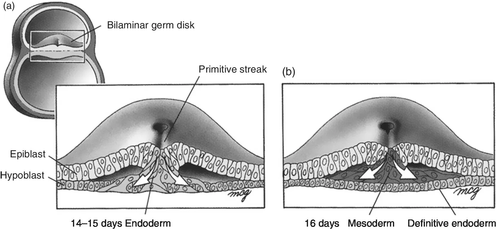
Around day 14, the embryonic bilaminar germ disk is composed of a layer of epiblast and a layer of hypoblast. At this time, a faint groove appears along the longitudinal midline of the germ disk that develops into a structure called the primitive streak [1]. Around day 15, epiblast cells near the primitive streak undergo a morphologic change and migrate through the primitive streak into the space between the epiblast and hypoblast in a process known as gastrulation (Fig. 1.1). Some of the ingressing epiblast cells invade the hypoblast, which is eventually replaced by a new layer of epiblast‐derived cells known as the definitive endoderm. Additional migrating epiblast cells occupy the space between the epiblast and the definitive endoderm to form a third layer of cells called the intraembryonic mesoderm (Fig. 1.1). As cells of the germinal disk migrate anteriorly to form a head process and lateral regions roll underneath to form an approximately cylindrical body shape, the endoderm is rolled into a tube that projects into the developing head region of the embryo surrounded by the mesoderm layer. This is the primitive digestive tube. The pancreas is specified by two separate outgrowths that arise on the dorsal and ventral surfaces of the primitive digestive tube. The epithelial cells of the pancreas originate from the interior lining of the primitive gut tube, which consists of a single layer of endoderm. A layer of mesenchyme, from which the muscle and connective tissue of the gastrointestinal organs are derived, surrounds the endoderm.
Figure 1.1 Germ disks sectioned through the region of the primitive streak, showing gastrulation. (a) On days 14 and 15, the ingressing epiblast cells replace the hypoblast to form the definitive endoderm. (b) The epiblast that ingresses on day 16 migrates between the endoderm and epiblast layers to form the intraembryonic mesoderm.
Source: Larsen 2001 [1]. Reproduced with permission of Elsevier.
The anterior regions of the endoderm form the foregut; regions posterior to the foregut form the midgut and hindgut. The most anterior regions of the foregut give rise to the esophagus and stomach. Just posterior to the foregut, the endoderm is continuous with the yolk sac, which extends outside the embryo, in a region known as the anterior intestinal portal. Endodermally derived cells close to the anterior intestinal portal specify the pancreas. The duodenum and liver are also specified by foregut endoderm in this region.
Thus, many gastrointestinal tissues are specified at the same time from a fairly restricted region of the gut endoderm. How are each of these organs specified in the appropriate anatomic location, and how do they differentiate properly into mature functional organs? The epithelial organs of the developing embryo originate as buds from the endoderm as the appropriate temporal and spatial cues are received. Thus, proper initiation and location of endodermally derived organs are regulated by the activation status of important signal transduction pathways involved in animal development, including the hedgehog, notch, and fibroblast growth factor signaling pathways.
Early Pancreatic Development
During the fourth week of gestation, two buds appear on the dorsal and ventral sides of the foregut near the anterior intestinal portal. These epithelial buds indicate the specification of the pancreas. These buds initially grow and differentiate independently, but later fuse to form a single organ. The anlage on the dorsal side, the dorsal pancreatic bud, appears first and gives rise to the dorsal pancreas. The cells of the dorsal pancreas will give rise to the head, body, and tail of the mature pancreas. The second pancreatic anlage appears shortly after the appearance of the dorsal pancreatic bud. This bud, which appears on the ventral side of the gut tube, is appropriately called the ventral pancreatic bud and develops into the ventral pancreas, which forms part of the head of the pancreas. Both pancreatic buds develop simultaneously, and the proliferating epithelial cells grow as projections into the surrounding mesenchymal tissue. During this time, the development of the intestines, and importantly the duodenum, continues. Rotation and asymmetric growth of the duodenum move the originally ventral part to a dorsal locati...