![]() Part One
Part One![]()
1
Structure and Function
In the following chapters of this book, we will deal with the examination and treatment of the human eye. As the majority of readers are expected to have an engineering or scientific background, we would like to provide a background in human ocular anatomy. This chapter shall also serve as an introduction and general reference for the more technical chapters. Of course, these chapters cannot claim to cover the entire anatomy and physiology of the eye, but they should be sufficient to gain an understanding of the interaction between the eye and the ophthalmic devices under consideration.
The human eye is a sophisticated sensory organ through which 80% of the sensory information we receive is processed. It is indeed our most important connection to the outside world. Any reduction or loss of vision means a major impairment of our quality of life.
In principle, the eye works like an artificial optical imaging system. To create an image, optical components focus light rays onto a photosensitive detector. But vision is more than just a projection of the surroundings onto a passive screen. The optical data is “preprocessed” by the photosensitive and neuronal tissue before it is sent to the brain for final “image analysis”. Even with efficient preprocessing, the eye sends 10 × more data to the brain than all other sensory organs altogether. To analyze this huge amount of information flow with almost no latency1), 30 different parts of the brain are involved simultaneously. During image processing, relevant information is filtered by recognition of known patterns.
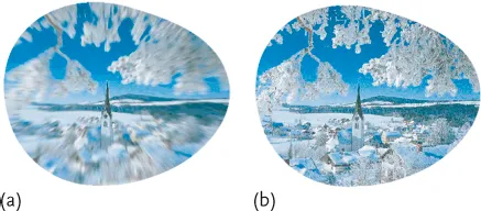
The brain’s important role for vision is illustrated in Figure 1.1. In Figure 1.1a, an animated image is shown as it would be directly projected onto photosensitive tissue. Here, a sharp image exists only in the center of the visual field. The eye now automatically changes the viewing direction in a fast manner and, for a moment, the margins of the image are sharply imaged as well. All the image segments are then merged by the brain so that the perceived field of sharp vision extends to the margins (Figure 1.1b).
Figure 1.1 Image processing by the brain. (a) Simulated “raw” image as it would be detected by photosensitive tissue. (b) Due to fast changes of the viewing direction and preprocessing of the “detected data”, the perceived field of clear vision is considerably extended. Taken with permission from [1].
We can trick the brain’s hard-wired processing algorithms by looking at visual illusions (Figure 1.2). It is an interesting and currently not fully answered question how much visual illusions are based purely on innate factors, or to what extent they are also based on experience and adaption.
Figure 1.2 Visual illusions which trick the image processing capability of the brain. (a) The checkerboard field A seems to be darker than B although the gray scales are equal in both cases. (b) The big dark dot at the top appears larger than the lower one, even though both dots are exactly the same size. (c) The circles seem to rotate. (d) Does the image show spirals? A closer look reveals that the structures are closed rings. (e) Fixate the pattern in the center. When moving your head, the circle seems to move independently from the background. When bringing your head closer, the pattern inside the circle seems to approach. (f) The lines appear to be bent although they are perfectly straight. See also [2].
1.1 Anatomy of the Human Eye
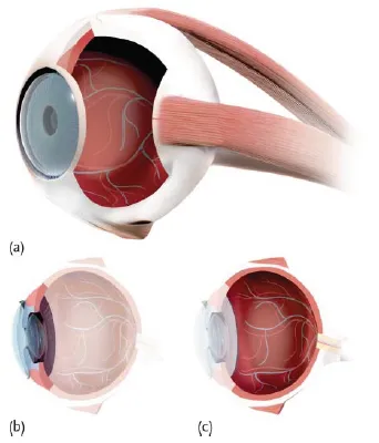
The human eye can be divided into the anterior and posterior segments (Figure 1.3b and c, respectively). The anterior segment (Figure 1.4) is the optical window to the environment. It mainly consists of optical components, such as the cornea, eye lens, and iris. The posterior segment of the eye (Figure 1.5) is referred to as the fundus. It is connected to the visual cortex of the brain via the optic nerve (Figure 1.6).
Figure 1.3 Anatomy of the human eye. (a) Oblique view of the human eye ball. (b) Side view of the eye with highlighted anterior segment. (c) Side view of the eye with highlighted posterior segment.
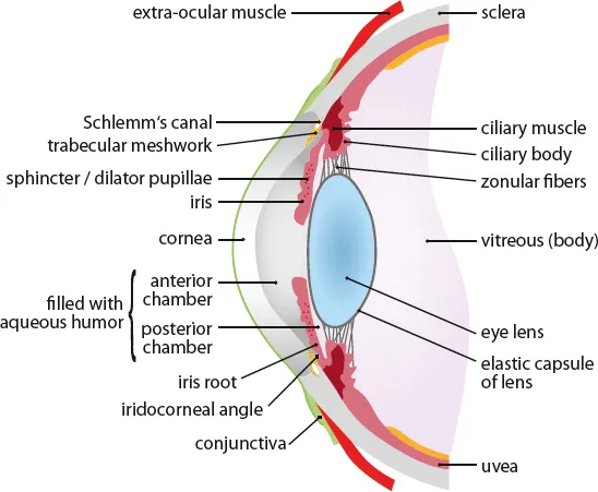
Figure 1.4 Scheme of the eye’s anterior segment (see also Figure 1.3b). The depicted eye components are not to scale.
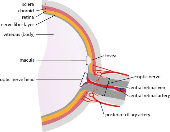
Figure 1.5 Scheme of the eye’s posterior segment (see also Figure 1.3c). The depicted eye components are not to scale.
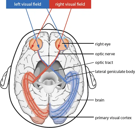
Figure 1.6 Transverse cross-section of the visual pathway including the primary visual cortex. The left and right areas of the retina are connected to different parts of the visual cortex. Hence, if one side of the visual cortex is impaired, the visual information of both eyes can still be used.
Sclera and cornea The spherical outer shell of the human eye consists of the white, opaque sclera. It serves as a mechanical support and protects the eye from injuries caused by mechanical force. The collagen fibers in the sclera are randomly distributed. Consequently, the incident light is strongly scattered (Section 9.2) so that the tissue appears white and opaque, which is why this part is also called “the whites of the eyes”.
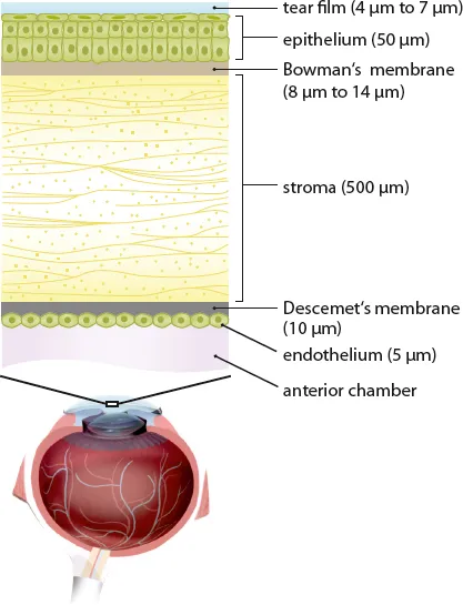
In the anterior segment of the eye, the sclera passes into the transparent cornea. The cornea is composed of multiple functional layers (Figure 1.7) and is covered by the 4–7 μm thick tear film. The tear film consists of a viscous, aqueous fluid that smooths the surface roughness of the corneal surface. A smooth surface reduces light scattering (Section 9.2), and thus improves the clarity of vision. The corneal epithelium is a 50 μm thick chemical barrier of the outer cornea which protects the eye against water, large molecules, and toxic substances. This is followed by Bowman’s membrane which is a thin layer (8–14 μm) above the 500 μm thick stroma. The stroma is composed of approximately 250 stacked collagen layers termed lamellae. Each lamella has a thickness of about 2 μm and contains ordered, cylindrical shaped collagen fibrils with diameters of 25–35 nm and a spacing of 20–50 nm [3]. Underneath the stroma we have the Descemet’s membrane (approximately 10 μm thick) which forms the basement layer of endothelial cells. The innermost corneal layer is the 5 μm thick endothelium. It is composed of hexagonal cells arranged in a honeycomb lattice and allows leakage of nutrients to the upper layers of the cornea. At the same time, the endothelium actively pumps water out of the cornea to keep it clear and transparent.
Figure 1.7 Detailed view of the corneal layer structure. The layer thicknesses are not to scale. Corresponding mean values are shown in parentheses. For reference, the cross-section of the whole eye is also shown. Adapted from [4].
Uvea, choroid, iris, and ciliary body The uvea forms the middle shell of the eye. In the posterior part of the eye (Figure 1.5), the uvea forms the so-called choroid, that is, a blood-rich tissue supplying nutrients to the retina (Section 1.2). The choroid has a total thickness of 350–450 μm. In the anterior segment (Figure 1.4), the uvea has evolved into the iris. In optical terms, the iris is an adjustable aperture stop whose diameter can be modified by two antagonistic muscles (sphincter and dilator pupillae). The hole of the iris is called the pupil. Note that the “anatomic pupil” does not correspond to the optical entrance or exit pupils (Sections 2.1.1 and A.1.4). The color of the iris depends on the amount of pigmentation in the anterior limiting layer of iris and stroma.
Between iris and choroid, the uvea has formed into the ciliary body which has two important functions. On the one hand, it produces the aqueous humor. On the other hand, it comprises the ciliary muscle which may relax the tension on the eye lens so that near vision is possible (Section 2.1.4).
Eye lens Similar to the cornea, the eye lens is a transparent tissue which contains no nerve fibers or blood vessels. The required nutrients are supplied by the aqueous humor, which is a clear fluid. The eye lens is embedded into an elastic capsule which is again attached to the ciliary body via zonular fibers. The capsule is composed of collagen and varies from 2–28 μm in thickness. The lens itself consists of an epithelial layer, which is only located in the anterior part of lens, and the lens fibers. The cells of the epithelium are located between the lens capsule and the outermost layer of lens fibers. Lens fibers form the bulk of the interior of the lens. They are long (up to 12 mm), thin, and transparent cells whose diameter ranges between 4 and 7 μm. The eye lens consists of two kinds of fiber. The inner core, the so-called nucleus, is formed by primary lens fibers. The nucleus is surrounded by the cortex which is formed by secondary lens fibers. The major purpose of the lens is the refractive change (i.e. accommodation; Section 2.1.4) to focus nearby objects.
Eye chambers The interior of the eye is divided into three chambers. The space between cornea and iris is called the anterior chamber, and between iris and eye lens we have the posterior chamber. The remaining space between lens and retina is referred to as the vitreous. The anterior and posterior chambers are filled with aqueous humor which contains nutrients for t...