![]()
Section 1
Introduction to Microorganisms and Antibacterial Chemotherapy
![]()
Chapter 1.1
Microorganisms
Key Points
- Bacteria can be classified according to their staining by the Gram stain (Gram positive, Gram negative, and mycobacteria) and their shape.
- Most bacterial (prokaryotic) cells differ from mammalian (eukaryotic) cells in that they have a cell wall and cell membrane, have no nucleus or organelles, and have different biochemistry.
- Bacteria can be identified by microscopy, or by using chromogenic (or fluorogenic) media or molecular diagnostic methods (e.g. real-time polymerase chain reaction (PCR)).
- Bacterial resistance to an antibacterial agent can occur as the result of alterations to a target enzyme or protein, alterations to the drug structure, and alterations to an efflux pump or porin.
- Antibiotic stewardship programmes are designed to optimise antimicrobial prescribing in order to improve individual patient care and slow the spread of antimicrobial resistance.
1.1.1 Classification
There are two basic cell types: prokaryotes and eukaryotes, with prokaryotes predating the more complex eukaryotes on earth by billions of years. Bacteria are prokaryotes, while plants, animals, and fungi (including yeasts) are eukaryotes. For our purposes in the remainder of this book, we will further subdivide bacteria into Gram positive, Gram negative, and mycobacteria (we will discuss prokaryotic cell shapes a little later).
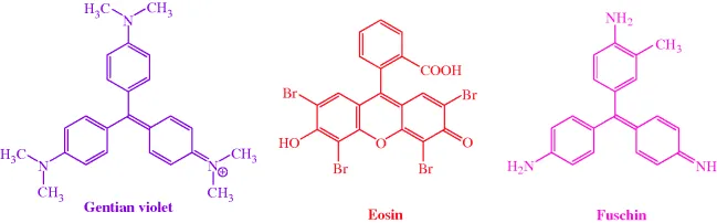
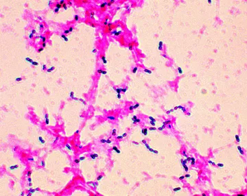
As you are probably already aware, we can use the Gram stain to distinguish between groups of bacteria, with Gram positive being stained dark purple or violet when treated with Gentian violet then iodine/potassium iodide (Figures 1.1.1 and 1.1.2). Gram negative bacteria do not retain the dark purple stain, but can be visualised by a counterstain (usually eosin or fuschin, both of which are red), which does not affect the Gram positive cells. Mycobacteria do not retain either the Gram stain or the counterstain and so must be visualised using other staining methods. Hans Christian Joachim Gram developed this staining technique in 1884, while trying to develop a new method for the visualisation of bacteria in the sputum of patients with pneumonia, but the mechanism of staining, and how it is related to the nature of the cell envelopes in these different classes of bacteria, is still unclear.
Some of the Gram positive and Gram negative bacteria, as well as some mycobacteria, which we shall encounter throughout this book, are listed in Table 1.1.1.
Table 1.1.1 Examples of Gram positive and Gram negative bacteria, and mycobacteria.
| Bacillus subtilis | Burkholderia cenocepacia | Mycobacterium africanum |
| Enterococcus faecalis | Citrobacter freundii | Mycobacterium avium complex (MAC) |
| Enterococcus faecium | Enterobacter cloacae | Mycobacterium bovis |
| Staphylococcus epidermis | Escherichia coli | Mycobacterium leprae |
| Staphylococcus aureus | Morganella morganii | Mycobacterium tuberculosis |
| Meticillin-resistant Staphylococcus aureus (MRSA) | Pseudomonas aeruginosa | |
| Streptococcus pyogenes | Salmonella typhimurium | |
| Listeria monocytogenes | Yersinia enterocolitica | |
1.1.2 Structure
The ultimate aim of all antibacterial drugs is selective toxicity – the killing of pathogenic1 bacteria (bactericidal agents) or the inhibition of their growth and multiplication (bacteriostatic agents), without affecting the cells of the host. In order to understand how antibacterial agents can achieve this desired selectivity, we must first understand the differences between bacterial (prokaryote) and mammalian (eukaryote) cells.
The name ‘prokaryote’ means ‘pre-nucleus’, while eukaryote cells possess a true nucleus, so one of the major differences between bacterial (prokaryotic) and mammalian (eukaryotic) cells is the presence of a defined nucleus (containing the genetic information) in mammalian cells, and the absence of such a nucleus in bacterial cells. Except for ribosomes, prokaryotic cells also lack the other cytoplasmic organelles which are present in eukaryotic cells, with the function of these organelles usually being performed at the bacterial cell membrane.
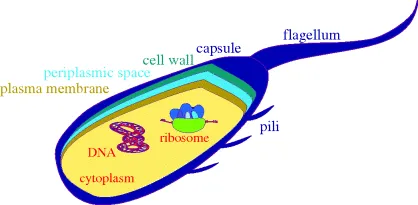
A schematic diagram of a bacterial cell is given in Figure 1.1.3, showing the main features of the cells and the main targets for antibacterial agents. As eukaryotic cells are much more complex, we will not include a schematic diagram for them here, and will simply list the major differences between the two basic cell types:
- Bacteria have a cell wall and plasma membrane (the cell wall protects the bacteria from differences in osmotic pressure and prevents swelling and bursting due to the flow of water into the cell, which would occur as a result of the high intracellular salt concentration). The plasma membrane surrounds the cytoplasm and between it and the cell wall is the periplasmic space. Surrounding the cell wall, there is often a capsule (there is also an outer membrane layer in Gram negative bacteria). Mammalian eukaryotic cells only have a cell membrane, whereas the eukaryotic cells of plants and fungi also have cell walls.
- Bacterial cells do not have defined nuclei (in bacteria the DNA is present as a circular double-stranded coil in a region called the ‘nucleoid’, as well as in circular DNA plasmids), are relatively simple, and do not contain organelles, whereas eukaryotic cells have nuclei containing the genetic information, are complex, and contain organelles,2 such as lysosomes.
- The biochemistry of bacterial cells is very different to that of eukaryotic cells. For example, bacteria synthesise their own folic acid (vitamin B9), which is used in the generation of the enzyme co-factors required in the biosynthesis of the DNA bases, while mammalian cells are incapable of folic acid synthesis and mammals must acquire this vitamin from their diet.
Whenever we discuss the mode of action of a drug, we will be focussing on the basis of any selectivity. As you will see from the section headings, we have classified antibacterial agents into those which target DNA (Section 2), metabolic processes (Section 3), protein synthesis (Section 4), and cell-wall synthesis (Section 5). In some cases, the reasons for antibacterial selectivity are obvious, for example mammalian eukaryotic cells do not have a peptidoglycan-based cell wall, so the agents we will discuss in Section 5 (which target bacterial cell-wall synthesis) should have no effect on mammalian cells. In other cases, however, the basis for selectivity is not as obvious, for example agents targeting protein synthesis act upon a process which is common to both prokaryotic and eukaryotic cells, so that in these cases selective toxicity towards the bacterial cells must be the result of a more subtle difference between the ribosomal processes in these cells.
We will now look at these antibacterial targets in detail, in preparation for our in-depth study of the modes of action of antibacterial agents and bacterial resistance in the remaining sections.
1.1.3 Antibacterial targets
1.1.3.1 DNA Replication
DNA replication is a complex process, during which the two strands of the double helix separate and each strand acts as a template for the synthesis of complementary DNA strands. This process occurs at multiple, specific locations (origins) along the DNA strand, with each region of new DNA synthesis involving many proteins (shown in italics below), which catalyse the individual steps involved in this process (Figure 1.1.4):
- The separation of the two strands at the origin to...