Atlas of Ear Diseases of the Dog and Cat
eBook - ePub

Atlas of Ear Diseases of the Dog and Cat

  1. English
  2. ePUB (mobile friendly)
  3. Available on iOS & Android
eBook - ePub

Atlas of Ear Diseases of the Dog and Cat

About this book

Bringing together a wealth of images of normal and diseased dog and cat ears, this is an indispensible diagnostic tool for the small animal veterinary practitioner seeing ear cases on a regular basis. This fully illustrated atlas covers the anatomy of the canine and feline ear, diagnostic techniques, a range of commonly seen diseases, and ear surgery.

Atlas of Ear Diseases of the Dog and Cat is one of the most complete picture references for this rapidly expanding branch of small animal medicine and surgery. It is an invaluable aid for general practitioners, as well as those specialising in dermatology, and serves as an effective revision aid for veterinary students and those studying for further qualifications in veterinary dermatology.

  • Includes over 400 high quality colour clinical images and clear line drawings
  • Images are accompanied by clear explanatory text throughout
  • Enables veterinarians to match cases seen in practice with photos supplied to aid diagnosis
  • Written by highly qualified specialist veterinary dermatologist and veterinary surgeon

Frequently asked questions

Yes, you can cancel anytime from the Subscription tab in your account settings on the Perlego website. Your subscription will stay active until the end of your current billing period. Learn how to cancel your subscription.
At the moment all of our mobile-responsive ePub books are available to download via the app. Most of our PDFs are also available to download and we're working on making the final remaining ones downloadable now. Learn more here.
Perlego offers two plans: Essential and Complete
  • Essential is ideal for learners and professionals who enjoy exploring a wide range of subjects. Access the Essential Library with 800,000+ trusted titles and best-sellers across business, personal growth, and the humanities. Includes unlimited reading time and Standard Read Aloud voice.
  • Complete: Perfect for advanced learners and researchers needing full, unrestricted access. Unlock 1.4M+ books across hundreds of subjects, including academic and specialized titles. The Complete Plan also includes advanced features like Premium Read Aloud and Research Assistant.
Both plans are available with monthly, semester, or annual billing cycles.
We are an online textbook subscription service, where you can get access to an entire online library for less than the price of a single book per month. With over 1 million books across 1000+ topics, we’ve got you covered! Learn more here.
Look out for the read-aloud symbol on your next book to see if you can listen to it. The read-aloud tool reads text aloud for you, highlighting the text as it is being read. You can pause it, speed it up and slow it down. Learn more here.
Yes! You can use the Perlego app on both iOS or Android devices to read anytime, anywhere — even offline. Perfect for commutes or when you’re on the go.
Please note we cannot support devices running on iOS 13 and Android 7 or earlier. Learn more about using the app.
Yes, you can access Atlas of Ear Diseases of the Dog and Cat by Sue Paterson,Karen Tobias in PDF and/or ePUB format, as well as other popular books in Medicine & Veterinary Medicine. We have over one million books available in our catalogue for you to explore.

Information

Year
2012
Print ISBN
9781405193269
eBook ISBN
9781118369098
Edition
1
CHAPTER 1
ANATOMY OF THE EAR
Karen Tobias
1.1 External Ear: Pinna and Ear Canal
1.2 Middle Ear of the Dog
1.3 Middle Ear of the Cat
1.4 Inner Ear

1.1 EXTERNAL EAR: PINNA AND EAR CANAL

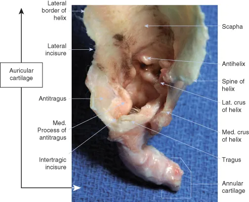
The pinna is the most prominent portion of the external ear (Fig. 1.1). It has an inner, concave surface and an outer, convex surface. In the standing ear, the concave surface forms a conchal cavity that is directed rostrally or laterally, while the convex surface faces medially or caudally. The distal tip of the pinna is called the apex, and the lateral and medial free margins of the pinna are called the helix (Fig. 1.2). The rostrolateral boundary of the distal portion of the ear canal is called the tragus. A notch caudal to the tragus, the intertragic incisure, separates it from the antitragus, which is a thin elongated piece of cartilage that extends up to the lateral margin of the helix at the cutaneous marginal pouch.
Figure 1.1 General anatomy of the pinna. The conchal cavity of the concave surface of the ear can be directed rostrally or laterally.
(Photo by Phil Snow, UTCVM) © 2012 The University of Tennessee.
c01f001
Figure 1.2 Concave surface of the right dog pinna. The antihelix and tragus form the boundaries of the ear canal opening.
(Photo by Phil Snow, UTCVM) © 2012 The University of Tennessee.
c01f002
The margins of the pinna are divided into medial, or rostral, and lateral, or caudal (see Fig. 1.1). These variations in directional description can make the anatomy very confusing.
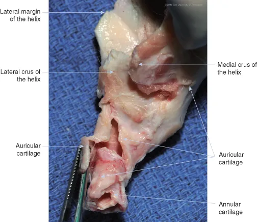
The external ear is composed of three cartilages: annular, auricular, and scutiform. The ear canal is formed proximally (near the skull) by the annular cartilage and distally (away from the skull) by the auricular cartilage, which fans out to form the pinna (Fig. 1.3).
Figure 1.3 Auricular and annular cartilage of the right ear of a dog, lateral view.
(Photo by Phil Snow, UTCVM) © 2012 The University of Tennessee.
c01f003
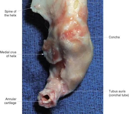
The auricular cartilage is divided into three sections: the scapha, the concha, and the tubus auris, or conchal tube (Fig. 1.4). Whereas the scapha is distally located and flattened, the concha is rolled into a trumpet shape to form the conchal cavity (Fig. 1.5). The scapha and concha are divided on the concave surface by the antihelix, a transverse cartilaginous fold.
Figure 1.4 Auricular and annular cartilage of the right ear, caudal view. The annular cartilage is nestled within the auricular cartilage, which forms the pinna and vertical ear canal. Note that the proximal portion of the auricular cartilage spirals inward as it bends.
(Photo by Phil Snow, UTCVM) © 2012 The University of Tennessee.
c01f004
Figure 1.5 Medial view of cartilage of the right ear. A portion of the auricular cartilage that forms the conchal tube has been elevated; underneath is another extension of auricular cartilage that wraps around the annular cartilage. Note that the ear canal is not a solid funnel: the auricular and annular tubes are each formed by overlapping flaps of cartilage that allow flexibility. Animals with severe otitis externa or conchal obstruction may develop periauricular abscesses from disruption of the fibrous connective tissue sheath surrounding either the tube flaps or the auricular-annular or annular-osseous junction.
(Photo by Phil Snow, UTCVM) © 2012 The University of Tennessee.
c01f005
The concha forms a funnel shape that thickens proximally as it becomes the conchal tube. The conchal tube forms the vertical ear canal. This canal is up to an inch (2.5 cm) deep and, as it progresses proximally towards the head, is directed ventrally, medially, and slightly rostral, spiralling inwards. It is partially surrounded along its proximal lateral border by the parotid salivary gland.
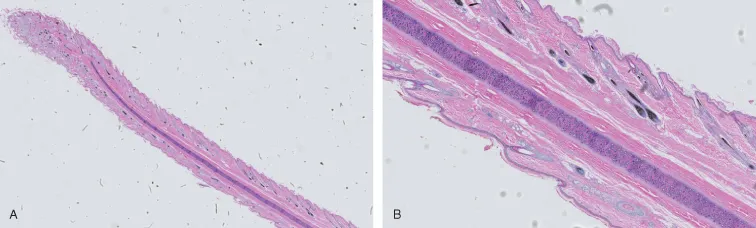
The annular cartilage is a separate, rolled, cartilaginous band that fits inside of the base of the conchal tube. It forms the horizontal ear canal, which runs medially toward the skull. In turn, the annular cartilage overlaps the osseous external acoustic meatus. Junctions of the auricular and annular cartilages and the annular cartilage and skull are connected by a fibrous tissue sheath. Because of these moveable joints, the auditory canal can be straightened during otoscopic examination. Epithelium lining the auricular and annular cartilage contains sebaceous and ceruminous glands and hair follicles (Fig. 1.6).
Figure 1.6 (A), Cross-section of the pinna of a dog. (B), Components including hyaline cartilage, muscle, and hair follicles are easily visible.
(Courtesy, UTCVM Virtual Microscope) © 2012 The University of Tennessee.
c01f006
Terminology for the ear canal varies within and amongst texts. Some authors consider the osseous extension of the skull that encompasses the tympanic membrane to be the external acoustic meatus or osseous external acoustic meatus, while others consider the external acoustic meatus to be the opening of the conchal tube at the level of the tragus and antihelix. The cartilaginous tube that extends from the meatus to the concha, which is a combination of conchal tube (auricular) and annular cartilage, is sometimes called the auditory canal.
A variety of muscles attach the ear rostrally, ventrally, or caudally to the head (Fig. 1.7); these muscles are innervated by the facial nerve. Some of these muscles are continuous with the cervical portion of the platysma. The plate-like, L-shaped scutiform cartilage, which is medial to the auricular cartilage, lies within the muscles that attach the auricular cartilage to the head (Fig. 1.8). By acting as a fulcrum, the scutiform cartilage improves mobility of the auricular cartilage.
Figure 1.7 Muscles of the canine ear and face: right lateral view...

Table of contents

  1. COVER
  2. DEDICATION
  3. TITLE PAGE
  4. COPYRIGHT PAGE
  5. FOREWORD
  6. PREFACE
  7. ACKNOWLEDGEMENTS
  8. CHAPTER 1 ANATOMY OF THE EAR
  9. CHAPTER 2 DIAGNOSTIC TECHNIQUES
  10. CHAPTER 3 ADVANCED DIAGNOSTIC TECHNIQUES
  11. CHAPTER 4 DISEASES OF THE EAR PINNA
  12. CHAPTER 5 CONDITIONS OF THE EAR CANAL
  13. CHAPTER 6 CONDITIONS OF THE MIDDLE EAR
  14. CHAPTER 7 DISEASES OF THE INNER EAR
  15. CHAPTER 8 SURGERY OF THE EAR
  16. INDEX