- English
- ePUB (mobile friendly)
- Available on iOS & Android
ABC of Emergency Radiology
About this book
Rapid acquisition and interpretation of radiographs, portable ultrasound (US) and computed tomography (CT) are now the mainstay of initial successful management of sick and traumatized patients presenting to Accident and Emergency Departments.
The ABC of Emergency Radiology is a simple and logical step-by-step guide on how to interpret radiographs, US and CT. It incorporates all the latest technological advances, including replacing plain radiographs with digital radiographs, changes in imaging protocols and the role of portable US and multidetector CT.
With over 400 illustrations and annotated radiographs, this thoroughly revised third edition provides more images, new illustrations, and new chapters on emergency US and CT that reflect current practice. Each chapter starts with radiological anatomy, standard and then additional views, a systematic approach to interpretation (ABC approach) and followed by a review of common abnormalities.
The ABC of Emergency Radiology is an invaluable resource for accident and emergency staff, trainee radiologists, medical students, nurses, radiographers and all medical personnel involved in the immediate care of trauma patients.
This title is also available as a mobile App from MedHand Mobile Libraries. Buy it now from https://itunes.apple.com/us/app/abc-emergency-radiology-3rd/id1055348839?ls=1&mt=8, https://www.medhand.com/products/abc-of-emergency-radiology or the https://www.medhand.com/products/abc-of-emergency-radiology.
Frequently asked questions
- Essential is ideal for learners and professionals who enjoy exploring a wide range of subjects. Access the Essential Library with 800,000+ trusted titles and best-sellers across business, personal growth, and the humanities. Includes unlimited reading time and Standard Read Aloud voice.
- Complete: Perfect for advanced learners and researchers needing full, unrestricted access. Unlock 1.4M+ books across hundreds of subjects, including academic and specialized titles. The Complete Plan also includes advanced features like Premium Read Aloud and Research Assistant.
Please note we cannot support devices running on iOS 13 and Android 7 or earlier. Learn more about using the app.
Information
Introduction: ABCs and Rules of Two
- Request the correct investigation
- Use a systematic approach to interpretation—ABCs
- Fundamental principles to avoid errors—Rules of two
- Always ASK for help—if in doubt!
- Plain to X rays (conventional or digital)
- Portable US
- MDCT
- MRI
MDCT—initial imaging modality of choice in the ED
| Head injuries/headaches or epilepsy | Skull X-ray (SXR) no longer done. CT head ± contrast |
| Facial injuries | MDCT with multiplanar reconstructions (MPR) and 3D are essential |
| Chest pain (suspected aortic aneurysm (AA), myocardial infarction (MI), pulmonary embolus (PE) or pneumothorax (Px)) | Triple rule out CT scan |
| Severe abdominal pain (obstruction) | CT has replaced abdominal X-ray (AXR) |
| Renal/ureteric colic | CT kidneys, ureters and bladder (KUB) has replaced Intravenous urogram (IVU) in ED |
| Suspected leaking abdominal aortic aneurysm (AAA) | CT has replaced US and AXR |
| Suspected gastrointestinal (GI) bleeding | Initially CT angiography instead of angiography |
| Major trauma (adults) | Whole body CT instead of chest X-ray (CXR), AXR and US |

Rules of Two

- Two views—one view is always one view too few
- Two abnormalities—if you see one abnormality, always look for a second
- Two joints—image the joint above
- Two sides—if not sure or difficult X ray, compare with other side
- Two views too many—CT (and rarely US) has replaced plain X rays in many clinical situations
- Two occasions—always compare with old films IF available
- Two visits—bring patient back for repeat examination
- Two opinions and two records—always ask a colleague if not sure and record findings
- Two specialists—always get your ED specialist and also a radiologist's opinion
- Two investigations—always consider whether US, CT or MRI would help in diagnosis
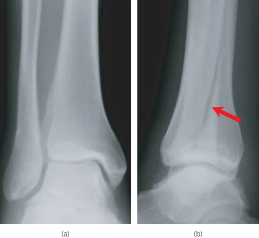
Rule 1—two views (‘One view is always one view too few’)
Table of contents
- Cover
- Series Page
- Title Page
- Copyright
- Dedication
- Contributors
- Preface
- Chapter 1: Introduction: ABCs and Rules of Two
- Chapter 2: Hand and Wrist
- Chapter 3: Elbow
- Chapter 4: Shoulder
- Chapter 5: Pelvis and Hip
- Chapter 6: Knee
- Chapter 7: Ankle and Foot
- Chapter 8: Head
- Chapter 9: Face
- Chapter 10: Cervical Spine
- Chapter 11: Thoracic and Lumbar Spine
- Chapter 12: Chest
- Chapter 13: Abdomen
- Chapter 14: Computed Tomography in Emergency Radiology
- Chapter 15: Emergency Ultrasound
- Chapter 16: Emergency Paediatric Radiology
- Chapter 17: Major Trauma
- Index
- Advertisement