Pituitary Disorders
eBook - ePub

Pituitary Disorders

Diagnosis and Management

Edward R. Laws, Shereen Ezzat, Sylvia L. Asa, Linda M. Rio, Lorin Michel, Robert Knutzen, Edward R. Laws, Shereen Ezzat, Sylvia L. Asa, Linda M. Rio, Lorin Michel, Robert Knutzen

  1. English
  2. ePUB (mobile friendly)
  3. Available on iOS & Android
eBook - ePub

Pituitary Disorders

Diagnosis and Management

Edward R. Laws, Shereen Ezzat, Sylvia L. Asa, Linda M. Rio, Lorin Michel, Robert Knutzen, Edward R. Laws, Shereen Ezzat, Sylvia L. Asa, Linda M. Rio, Lorin Michel, Robert Knutzen

Book details
Book preview
Table of contents
Citations

About This Book

Do you want to be up to date on the latest concepts of diagnosis and treatment of patients suffering from disorders of the pituitary gland?

Are you looking for an expert guide to the best clinical management?

If so, this is the book for you, providing a full analysis of pituitary disorder management from acromegaly to Addison's Disease; from Cushing's Disease to hypopituitarism; from hormone disorders to hormone replacement.

Well-illustrated throughout, and with contributions from leading specialists in pituitary disease, inside you'll find comprehensive and expert coverage, including:

  • Diagnosing pituitary disease
  • Management options for each disorder
  • Complications that can occur
  • Psychological and psychosocial effects of pituitary disease
  • What outcomes you and your patients can expect over the long term
  • Current research and clinical trials related to pituitary disease

Pituitary Disorders: Diagnosis and Management is the perfect clinical tool for physicians and health care providers from many related disciplines, and an essential companion for the best quality management of pituitary patients.

Frequently asked questions

How do I cancel my subscription?
Simply head over to the account section in settings and click on “Cancel Subscription” - it’s as simple as that. After you cancel, your membership will stay active for the remainder of the time you’ve paid for. Learn more here.
Can/how do I download books?
At the moment all of our mobile-responsive ePub books are available to download via the app. Most of our PDFs are also available to download and we're working on making the final remaining ones downloadable now. Learn more here.
What is the difference between the pricing plans?
Both plans give you full access to the library and all of Perlego’s features. The only differences are the price and subscription period: With the annual plan you’ll save around 30% compared to 12 months on the monthly plan.
What is Perlego?
We are an online textbook subscription service, where you can get access to an entire online library for less than the price of a single book per month. With over 1 million books across 1000+ topics, we’ve got you covered! Learn more here.
Do you support text-to-speech?
Look out for the read-aloud symbol on your next book to see if you can listen to it. The read-aloud tool reads text aloud for you, highlighting the text as it is being read. You can pause it, speed it up and slow it down. Learn more here.
Is Pituitary Disorders an online PDF/ePUB?
Yes, you can access Pituitary Disorders by Edward R. Laws, Shereen Ezzat, Sylvia L. Asa, Linda M. Rio, Lorin Michel, Robert Knutzen, Edward R. Laws, Shereen Ezzat, Sylvia L. Asa, Linda M. Rio, Lorin Michel, Robert Knutzen in PDF and/or ePUB format, as well as other popular books in Medizin & Endokrinologie & Stoffwechsel. We have over one million books available in our catalogue for you to explore.

Information

Year
2013
ISBN
9781118559376
SECTION 1
Overview
CHAPTER 1
The Endocrine System
Sylvia L. Asa and Shereen Ezzat
University of Toronto, Toronto, ON, Canada

Normal Development and Structure

The endocrine system is composed of cells and organs that have, as their primary function, the production and secretion of hormones. They are generally classified into three broad categories: peptide hormone-producing, steroid hormone-producing, and thyroid hormone-producing.

Peptide Hormone-Producing Cells

The majority of endocrine cell types produce peptide hormones. This group of endocrine cells have a characteristic morphology that is called “neuroendocrine” because of its similarity to neural cells [1]. They have sufficient neural differentiation structurally and functionally that they have been called “paraneurons.” Historically they were classified as the APUD (amine precursor uptake and decarboxylation) system. It was previously suggested that they derive embryologically from the neural crest, but this has not been proven for all members of this group of cells, many of which arise from the primitive endoderm. Nevertheless, functionally they act as neuron-like cells; they secrete peptides that are often also produced by neurons. In fact, endocrine cells and neurons are like conventional and wireless communication: neurons produce messengers that are released at synapses and activate receptors in physically adjacent cells, rather like conventional wiring, whereas neuroendocrine cells produce the same types of messengers but release them into the bloodstream to activate cells throughout the body, analogous to wireless messages that do not rely on physical contact for communication.
These cells aggregate into classical endocrine organs, the pituitary, parathyroid, and adrenal medulla, and are also found singly and in small clusters of the dispersed endocrine system, scattered within other organs, such as the calcitonin-secreting C cells of the thyroid, and the endocrine cells of the lung, gut, and pancreas. The wide array of peptide hormones they produce is essential for regulation of most metabolic and reproductive functions.

Steroid Hormone-Producing Cells

Steroid hormone-producing cells are primarily found in the adrenal cortex and the gonads. They also have a distinct morphology that reflects their primary function of conversion of cholesterol into the various mineralocorticoid, glucocorticoid, an­­drogenic, and estrogenic hormones. They are of mesodermal origin arising from the coelomic epithelium that gives rise to the adrenal and the genital ridge.

Thyroid Hormone-Producing Cells

Thyroid hormone-producing cells are modified epithelial cells derived from the oral endoderm that invaginate from the base of tongue. They are specifically involved in the synthesis of thyroglobulin and its iodination to form thyroid hormones.

Endocrine Regulation

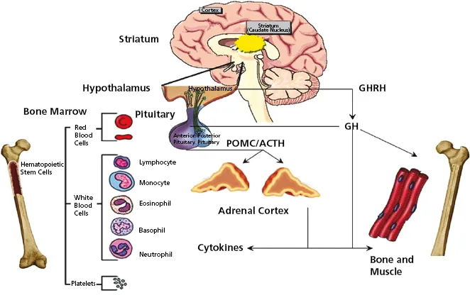
The endocrine system is tightly regulated by hormones that stimulate target endocrine cells and in turn respond to suppression by the products of their targets. The hypothalamic–pituitary axis is the central regulatory system (Figure 1.1, left). Through this axis, there is central regulation of growth, adrenal and thyroid metabolic function, reproduction, and breast development and function. Direct and indirect mechanisms involved in this system regulate immunity and emotional status (Figure 1.2). The hypothalamus and posterior pituitary also regulate salt and water homeostasis as well as lactation (Figure 1.1, right). A separate axis regulates nutrient metabolism through the pancreatic islets and gut (Figure 1.1, right). There is evidence that this too is under central control, but mainly by regulation of appetite [2]. Finally, the sympathetic and parasympathetic nervous systems regulate endocrine function through the adrenal medulla and paraganglia [3].
Figure 1.1. The endocrine system is composed of cells, groups of cells, and organs that have as their main function the production of hormones that regulate homeostasis throughout the body. The hypothalamus is the central mediator of the system where it integrates neuronal input with feedback from target organs. Via the posterior pituitary, the hypothalamus regulates salt and water resorption in the kidney through vasopressin; it also regulates breast lactation through oxytocin. Hypothalamic control of the anterior pituitary regulates thyroid, adrenal, and gonadal function, as well as growth of bone and muscle through growth hormone-mediated liver production of IGF-1. The pancreas and gut represent an independent endocrine regulatory system that modulates nutrient absorption and utilization.
(Illustration by Sonia Chang.)
web_c1-fig-0001
Figure 1.2. Ikaros is expressed in restricted sites throughout the neuroendocrine and hematopoietic systems. In the brain, the highest expression is in the medium spiny neurons of the striatum where the loss of function results in neurobehavioral changes characterized by an anti-depressant phenotype. In the hypothalamus, the median eminence of GHRH-containing neurons colocalize with Ikaros expression. Loss of Ikaros severely diminishes GHRH production and consequently GH and IGF-1 activation. In the anterior pituitary, Ikaros is expressed in POMC-producing corticotrophs that govern the ACTH/adrenocortical axis. Ikaros is also expressed in the somatotrophs where it plays a direct inhibitory role. In the hematopoietic system, peak expression of Ikaros is in stem cells where it directs lymphoid lineage commitment. The multidimensional actions of Ikaros serve to sort and integrate diverse signals to regulate neuroendocrine–immune interactions through direct and indirect mechanisms. (Source: Ezzat S, Asa SL. The emerging role of the Ikaros stem cell factor in the neuroendocrine system. Journal of Molecular Endocrinology 2008; 41:45–51.)
web_c1-fig-0002

Endocrine Pathology

This review will provide information on pathologies of the endocrine system with a focus on the role of the pituitary in endocrine homeostasis. In general, endocrine homeostasis is altered when there is hypofunction, resulting in hormone deficiencies, or hyperfunction, due to hormone excess. Endocrine deficiencies can result from many pathological processes, as discussed later. Hormone excess is almost always due to hyperplasia or neoplasia.

Endocrine Deficiencies

Deficiency of endocrine function can be attrib­uted to several types of pathological processes. Fortunately, most hormone deficiencies can be treated with hormone replacement regimens.

Isolated Hormonal Deficiencies or Resistance

These are usually caused by genetic mutations that interrupt the production of hormones, their receptors, or the enzymes required for their actions. The most common isolated hormone deficiency is congenital hypothyroidism [4], which can result in dyshormonogenetic goiter and cretinism, but in many countries complications are prevented by screening programs of neonates that lead to thy­­roid hormone replacement. Congenital adrenal hyperplasia is a spectrum of disorders due to a defect in one of the five enzymatic steps involved in steroid synthesis [5]; 90–95% of cases are caused by deficiency of 21-hydroxylase, resulting in marked elevation of 17-hydroxyprogesterone and male hormone excess at the price of diminished glucocorticoid reserves.
Isolated pituitary hormone deficiency most commonly involves growth hormone [6]. Rare examples of thyroid hormone receptor [7]or glucocorticoid receptor resistance [8] result in similar clinical manifestations as loss of hormone itself.

Tissue Destruction

Tissue destruction resulting in hormone deficiencies is another major cause of hormone deficiency. Tissue destruction can be the result of surgery, or may be caused by pressure or infiltration of the organ or cells by cancer or inflammation. There are many examples of each of these types of endocrine hypofunction. The most common iatrogenic hormone deficiency is hypoparathyroidism following thyroid surgery. In the pituitary, compression of normal tissue by cysts or tumors can result in hypopituitarism; tissue resection at the time of surgery can exacerbate hypopituitarism.
Inflammatory conditions can cause endocrine dysfunction, although acute and chronic infections rarely cause endocrine deficiencies in the Western world. In the sella turcica [9] this can happen in association with sphenoid sinus infection, cavernous sinus thrombosis, by spread of otitis media mastoiditis or peritonsillar abscess, or rarely by vascular seeding of distant or systemic infection by a wide variety of infectious agents, including fungi, mycobacteria, bacteria, and spirochetes. Other causes of secondary hypophysitis include sar­coidosis, vasculitides such as Takayasu’s disease and Wegener’s granulomatosis, Crohn’s disease, Whipple’s disease, ruptured Rathke’s cleft cyst, necrotizing adenoma, and meningitis. Complications of AIDS may also involve endocrine tissues, including the pituitary gland; involvement is usually infectious in nature (including Pneumocystis jirovecii, toxoplasmosis, and cytomegalovirus) and results in acute or chronic inflammation with necrosis.
Autoimmune endocrine disorders are a significant cause of hormone deficiency. Examples in­­clude type 1 diabetes mellitus due to autoimmune destruction of insulin-producing cells of the pancreatic islets and hypothyroidism due to the various forms of chronic lymphocytic thyroiditis including Hashimoto’s thyroiditis. Autoimmune inflammation has been described in almost every endocrine tissue. Most of the rare variants are associated with polyendocrine autoimmune syndromes that predispose individuals to immune destruction of endocrine and nonendocrine cells in multiple tissues, both endocrine and nonendocrine, the latter including melanocytes of the skin (resulting in vitiligo) and parietal cells of the stomach (resulting in pernicious anemia). The autoimmune polyendocrine syndrome type 1 (APS1) is the most well understood of these disorders, since its pathogenesis has been recently elucidated. This monogenic autoimmune syndrome is caused by mutations in the autoimmune regulator (AIRE) gene on chromosome 21 that encodes a nuclear protein involved in transcriptional processes and the regulation of self-antigen expression in thymus [10]. High-titer autoantibodies toward intracellular enzymes are a hallmark of APS1 and serve as diagnostic markers and predictors for disease manifestations.
In the pituitary, lymphocytic hypophysitis has been attributed to autoimmunity [9]. The disease is associated with other endocrine autoimmune phenomena and forms part of APS1; a tudor domain-containing protein 6 (TDRD6) was identified as the target of a putative autoantibody in APS1 patients and in patients with growth hormone (GH) deficiency, and is expressed in pituitary [11], but it remains to be proven if this is the causative antigen. The association of the classical form of lymphocytic hypophysitis with pregnancy may be attributed to hyperplasia of ...

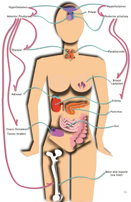
Table of contents

Citation styles for Pituitary Disorders

APA 6 Citation

[author missing]. (2013). Pituitary Disorders (1st ed.). Wiley. Retrieved from https://www.perlego.com/book/1000737/pituitary-disorders-diagnosis-and-management-pdf (Original work published 2013)

Chicago Citation

[author missing]. (2013) 2013. Pituitary Disorders. 1st ed. Wiley. https://www.perlego.com/book/1000737/pituitary-disorders-diagnosis-and-management-pdf.

Harvard Citation

[author missing] (2013) Pituitary Disorders. 1st edn. Wiley. Available at: https://www.perlego.com/book/1000737/pituitary-disorders-diagnosis-and-management-pdf (Accessed: 14 October 2022).

MLA 7 Citation

[author missing]. Pituitary Disorders. 1st ed. Wiley, 2013. Web. 14 Oct. 2022.