Practical Flow Cytometry in Haematology
eBook - ePub

Practical Flow Cytometry in Haematology

100 Worked Examples

  1. English
  2. ePUB (mobile friendly)
  3. Available on iOS & Android
eBook - ePub

Practical Flow Cytometry in Haematology

100 Worked Examples

About this book

The analysis of blood, bone marrow and tissue fluid specimens requires a multi-faceted approach with the integration of scientific data from a number of disciplines. No single discipline can operate in isolation or errors will occur. Flow cytometry is in a privileged position in that it can provide rapid analysis of specimens and it is often the first definitive investigation to produce results and help formulate a working diagnosis.

This companion text to Practical Flow Cytometry in Haematology Diagnosis contains 100 worked examples drawn from real clinical cases presenting to the authors' institution. Cases are illustrated with peripheral blood and bone marrow cytology, tissue pathology and cytogenetic and molecular data, which are integrated to generate, where appropriate, a diagnosis based on the WHO Classification of Tumours of Haematopoietic and Lymphoid Tissues. The spectrum of clinical cases includes adult and paediatric patients, and both neoplastic and reactive disorders. The cases appear in no particular order to challenge the reader to make their own diagnosis.
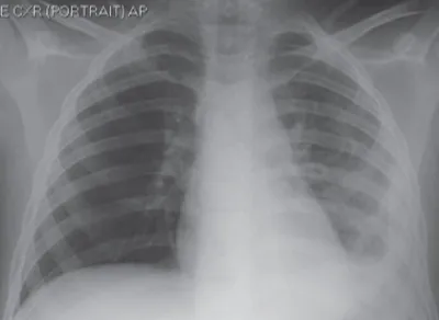
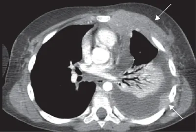
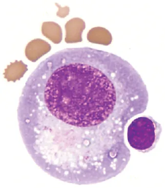
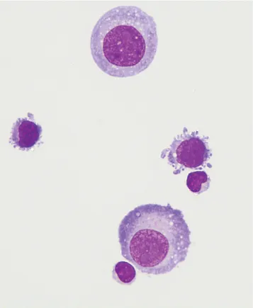
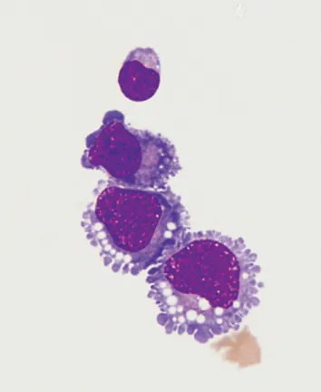
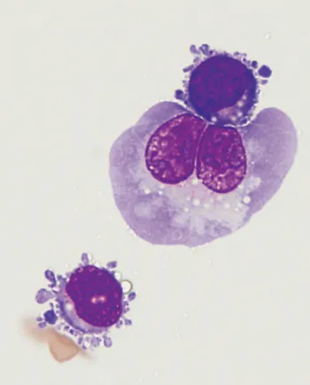
The reader will review May?Grรผnwald?Giemsa (MGG)-stained films of peripheral blood and bone marrow aspirates presented alongside flow cytometric data and haematoxylin and eosin (H&E)-stained bone marrow and other tissue biopsy sections. Immunohistochemistry is used to further clarify the tissue lineage and cell differentiation. Cytogenetic studies using metaphase preparations are used to identify translocations and chromosome gains and losses whilst interphase fluorescence in situ hybridisation (FISH) studies and polymerase chain reaction (PCR) are used to identify gene fusions, gene rearrangements and deletions. Each case concludes with a discussion of the features that are important to making a diagnosis. The cases are also listed according to disease classification in the appendix so that the text can also be used as a reference.

Practical Flow Cytometry in Haematology: 100 Worked Examples:

  • Provides a practical, example-based resource for flow cytometry
  • Demonstrates how flow cytometry results should be interpreted and applied to optimize patient care
  • Includes both malignant and benign conditions
  • Can be used in conjunction with Practical Flow Cytometry in Haematology Diagnosis, by the same author team (ISBN 9780470671207)

Practical Flow Cytometry in Haematology: 100 Worked Examples is ideal for practicing haematologists and histopathologists with an interest in haematopathology, but particularly directed at trainee haematologists and scientists preparing for FRCPath and related examinations.

Frequently asked questions

Yes, you can cancel anytime from the Subscription tab in your account settings on the Perlego website. Your subscription will stay active until the end of your current billing period. Learn how to cancel your subscription.
At the moment all of our mobile-responsive ePub books are available to download via the app. Most of our PDFs are also available to download and we're working on making the final remaining ones downloadable now. Learn more here.
Perlego offers two plans: Essential and Complete
  • Essential is ideal for learners and professionals who enjoy exploring a wide range of subjects. Access the Essential Library with 800,000+ trusted titles and best-sellers across business, personal growth, and the humanities. Includes unlimited reading time and Standard Read Aloud voice.
  • Complete: Perfect for advanced learners and researchers needing full, unrestricted access. Unlock 1.4M+ books across hundreds of subjects, including academic and specialized titles. The Complete Plan also includes advanced features like Premium Read Aloud and Research Assistant.
Both plans are available with monthly, semester, or annual billing cycles.
We are an online textbook subscription service, where you can get access to an entire online library for less than the price of a single book per month. With over 1 million books across 1000+ topics, weโ€™ve got you covered! Learn more here.
Look out for the read-aloud symbol on your next book to see if you can listen to it. The read-aloud tool reads text aloud for you, highlighting the text as it is being read. You can pause it, speed it up and slow it down. Learn more here.
Yes! You can use the Perlego app on both iOS or Android devices to read anytime, anywhere โ€” even offline. Perfect for commutes or when youโ€™re on the go.
Please note we cannot support devices running on iOS 13 and Android 7 or earlier. Learn more about using the app.
Yes, you can access Practical Flow Cytometry in Haematology by Mike Leach,Mark Drummond,Allyson Doig,Pam McKay,Bob Jackson,Barbara J. Bain in PDF and/or ePUB format, as well as other popular books in Medicine & Hematology. We have over one million books available in our catalogue for you to explore.

Information

Year
2015
Print ISBN
9781118747032
eBook ISBN
9781118746899
Edition
1
Subtopic
Hematology

Case 1

An 11-year-old boy was admitted with a short history of fever, sweats, dyspnoea and left chest discomfort. There was no past history of note. Examination identified features of a left pleural effusion. There was also a tender swelling of the left anterior chest in the upper pectoral region and palpable cervical lymphadenopathy. The liver and spleen were not palpable.

Laboratory investigations

FBC and blood film: normal
U&Es, LFTs: normal. LDH was 1460 U/L.

Imaging

The CXR showed opacification and loss of aeration of the left hemithorax in keeping with a pleural effusion (Figure 1.1). CT imaging confirmed this but in addition identified a left pleural-based mass, abnormal soft tissue in the left pectoral muscles (arrows, Figure 1.2) and cervical lymphadenopathy. In addition, there was collapse/consolidation of the lower left lung, creating the appearance of an air bronchogram. A core biopsy of a cervical node was taken and the pleural effusion was aspirated for analysis.
c01f001
Figure 1.1 CXR.
c01f002
Figure 1.2 CT.

Flow cytometry

The pleural fluid cell count was 0.98 ร— 109/L. A cytospin preparation showed three distinct cell types: a small mature lymphoid population in keeping with reactive lymphocytes, an intermediate sized/large sized lymphoid population and a large cell population with pleomorphic morphology and blue cytoplasm (Figures 1.3โ€“1.6). The cells with the abundant cytoplasm (Figures 1.3 and 1.4) and the single binucleate cell (Figure 1.6) are reactive mesothelial cells. The cells with the cytoplasmic blebs (Figures 1.4โ€“1.6) are the disease cells, which were the subsequent focus for immunophenotyping studies.
c01f003
Figure 1.3 MGG, ร—500.
c01f004
Figure 1.4 MGG, ร—500.
c01f005
Figure 1.5 MGG, ร—500.
c01f006
Figure 1.6 MGG, ร—500.
By applying a blast gate to the suspected malignant cells in the FSC/SSC analysis (Figure 1.7), they were shown to express CD45bright (Figure 1.8), CD2 (Figure 1.9), cCD3 [whilst surface CD3 was negative apart from a few reactive T cells (Figure 1.8)], partial CD7 (Figure 1.10) and CD13. Othe...

Table of contents

  1. Cover
  2. Title Page
  3. Copyright
  4. Table of Contents
  5. Preface
  6. Acknowledgement
  7. List of Abbreviations
  8. Technical Notes
  9. Laboratory Values
  10. Case 1
  11. Case 2
  12. Case 3
  13. Case 4
  14. Case 5
  15. Case 6
  16. Case 7
  17. Case 8
  18. Case 9
  19. Case 10
  20. Case 11
  21. Case 12
  22. Case 13
  23. Case 14
  24. Case 15
  25. Case 16
  26. Case 17
  27. Case 18
  28. Case 19
  29. Case 20
  30. Case 21
  31. Case 22
  32. Case 23
  33. Case 24
  34. Case 25
  35. Case 26
  36. Case 27
  37. Case 28
  38. Case 29
  39. Case 30
  40. Case 31
  41. Case 32
  42. Case 33
  43. Case 34
  44. Case 35
  45. Case 36
  46. Case 37
  47. Case 38
  48. Case 39
  49. Case 40
  50. Case 41
  51. Case 42
  52. Case 43
  53. Case 44
  54. Case 45
  55. Case 46
  56. Case 47
  57. Case 48
  58. Case 49
  59. Case 50
  60. Case 51
  61. Case 52
  62. Case 53
  63. Case 54
  64. Case 55
  65. Case 56
  66. Case 57
  67. Case 58
  68. Case 59
  69. Case 60
  70. Case 61
  71. Case 62
  72. Case 63
  73. Case 64
  74. Case 65
  75. Case 66
  76. Case 67
  77. Case 68
  78. Case 69
  79. Case 70
  80. Case 71
  81. Case 72
  82. Case 73
  83. Case 74
  84. Case 75
  85. Case 76
  86. Case 77
  87. Case 78
  88. Case 79
  89. Case 80
  90. Case 81
  91. Case 82
  92. Case 83
  93. Case 84
  94. Case 85
  95. Case 86
  96. Case 87
  97. Case 88
  98. Case 89
  99. Case 90
  100. Case 91
  101. Case 92
  102. Case 93
  103. Case 94
  104. Case 95
  105. Case 96
  106. Case 97
  107. Case 98
  108. Case 99
  109. Case 100
  110. Antibodies Used in Immunohistochemistry Studies
  111. Flow Cytometry Antibodies
  112. Molecular Terminology
  113. Classification of Cases According to Diagnosis
  114. Index
  115. End User License Agreement