- Combines new and influential research, along with articulate overviews of the key topics in theoretical and applied areas of speech communication
- Accessibly structured into five major sections covering: experimental phonetics; biological perspectives; modelling speech production and perception; linguistic phonetics; and speech technology
- Includes nine entirely new chapters on topics such as phonetic notation and sociophonetics, speech technology, biological perspectives, and prosody
- A streamlined and re-oriented structure brings all contributions up-to-date with the latest research, whilst maintaining the features that made the first edition so useful
eBook - ePub
The Handbook of Phonetic Sciences
- English
- ePUB (mobile friendly)
- Available on iOS & Android
eBook - ePub
The Handbook of Phonetic Sciences
About this book
Thoroughly revised and updated, the second edition of The Handbook of Phonetic Sciences provides an authoritative account of the key topics in both theoretical and applied areas of speech communication, written by an international team of leading scholars and practitioners.
Trusted by 375,005 students
Access to over 1.5 million titles for a fair monthly price.
Study more efficiently using our study tools.
Information
Part I
Experimental Phonetics
1
Laboratory Techniques for Investigating Speech Articulation
This chapter discusses current laboratory techniques that measure the oral vocal tract during speech. The focus is on instruments that measure the articulators directly and indirectly. Indirect measurements come from instruments that are remote from the structures of interest such as imaging techniques. Direct measurements come from instruments that contact the structures of interest, such as, point-tracking devices and electropalatography. Although some references are made to current research using each instrument, to indicate its applications and strengths, the list of studies is not comprehensive as the goal is to explain the instrument.
Measuring the vocal tract is a challenging task because the articulators differ widely in location, shape, structural composition, and speed and complexity of movement. First, there are large differences in tissue consistency between soft tissue structures (tongue, lips, velum) and hard tissue structures (jaw, palate), which result in substantially different movement complexity. In other words, the fluid deformation of the soft structures and the rigid movements of the bones need different measurement strategies. Second, measurement strategies must differ between structures visible to superficial inspection, such as the lips, and structures deep within the oral cavity, such as the velum. Third, articulator rates of motion vary, so that an instrument with a frequency response appropriate for the slow-moving jaw will be too slow for the fast-moving tongue tip. The final and perhaps most important measurement complication is the interaction among articulators. Some articulatory behaviors are highly correlated, and distinguishing the contributions of each player can be quite difficult. The most dramatic example of this is the tongue–jaw system. It is clear that jaw height is a major factor in tongue tip height. However, the coupling of these two structures becomes progressively weaker as one moves posteriorly, until in the pharynx, tongue movement is only minimally coupled to jaw movement if at all. Thus, trying to measure the contribution of the jaw to tongue movement becomes a difficult task.
It is difficult to devise a transducer that can be inserted into the mouth, which will not in some way distort the speech event. Thus, the types of instruments used in the vocal tract need to be unobtrusive, such as by resting passively against a surface (e.g., electropalatography), by being small and positioned on noncontact surfaces (e.g., pellet tracking systems), or by not entering the vocal tract at all (e.g., imaging techniques).
Instruments that enter the oral cavity must meet certain criteria. They need to be unaffected by temperature change, moisture, or air pressure. Affixatives must be unaffected by moisture, nontoxic, able to stick to expandable, moist surfaces, and must be removable without tearing the surface tissue. Devising instruments that are noninvasive, unobtrusive, meet the above criteria, and still measure one or more components of the speech event is so difficult that most researchers prefer to study the speech wave and infer physiological events from it. However, since those inferences are based on, and refined by, physiological data, it is critical to add new physiological databases, lest models of the vocal tract and our understanding of speech production stagnate.
In recent times, physiological measurements have improved at an extraordinary pace. Imaging techniques are revolutionizing the way we view the vocal tract by providing recognizable images of structures deep within the pharynx. They also provide information on local tissue movement and control strategies. Point-tracking systems and palatographic measurements have transformed our ideas about coarticulation by revealing inter-articulator relationships that could only in the past be addressed theoretically. Applications to linguistics and rehabilitation are now ongoing. This chapter considers indirect measurements, that is, imaging techniques, and direct measurements such as point-tracking techniques, and tongue–palate measurement devices
1 Imaging Techniques
The internal structures of the vocal tract are difficult to measure without impinging upon normal movement patterns. Imaging techniques overcome that difficulty because they register internal movement without directly contacting the structures. Four well-known imaging techniques have been applied to speech research: X-ray, computed tomography (CT), magnetic resonance imaging (MRI), and ultrasound. Imaging systems provide recordings of the entire structure, rather than single points on the structure.
1.1 X-ray
X-ray is the most well known of the imaging systems. It is important because it was the first widely used imaging system and most of our historical knowledge about the pharyngeal portion of the vocal tract came from X-ray data. To make a lateral X-ray image, an X-ray beam is projected from one side of the head through all the tissue, and recorded onto a plate on the other side. The resulting image shows the head from front to back and provides a lengthwise view of the tongue. A frontal or anterior–posterior (AP) X-ray is made by projecting the X-ray beam from the front of the head through to the back of the head and recording the image on a plate behind the head. The resulting images provide a cross-sectional view of the oral cavity. Prior to the advent of MRI considerable research was done using X-ray imaging. More recent X-ray studies are based on archival databases.
X-ray data have contributed to many aspects of speech production research. Many vocal tract models are based on X-rays (cf. Fant, 1965; Mermelstein, 1973; Harshman et al., 1977; Wood, 1979; Hashimoto & Sasaki, 1982; Maeda, 1990). X-rays have also been used to study normal speech production (Kent & Netsell, 1971; Kent, 1972; Kent & Moll, 1972), nonspeech motions (Kokawa et al., 2006), motor control strategies (Lindblom et al., 2002; Iskarous, 2005), language differences (cf. Gick, 2002b; Gick et al., 2004), and speech disorders (Subtelny et al., 1989; Tye-Murray, 1991).
Usually soft tissue structures such as the tongue are difficult to measure with X-rays, because the beam records everything in its path including teeth, jaw, and vertebrae. These strongly imaged bony structures obscure the fainter soft tissue. Another limitation of X-ray is that unless a contrast medium is used to mark the midline of the tongue, it is difficult to tell if the visible edge is the midline surface of the tongue or a lateral edge. This is particularly problematic during speech, because the tongue is often grooved or arched. Finally, the potential hazards of overexposure have reduced the collection of large quantities of X-ray data. There is, however, public availability of archival X-ray databases for research use. One such database (Munhall et al., 1994a, 1994b) was compiled by Advanced Technologies Research Laboratories, Kyoto, and is available from http://psyc.queensu.ca/∼munhallk/05_database.htm.
1.2 Tomography
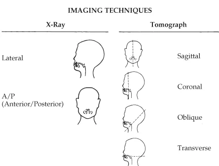
Tomography is a fundamentally different imaging method from projection X-ray in that it records slices of tissue. Three tomographic techniques used in speech research are Computed Tomography, Magnetic Resonance Imaging, and Ultrasound Imaging. These slices are made by projecting a thin, flat beam through the tissue in one of four planes: sagittal, coronal, oblique, and transverse (see Figure 1.1). The mid-sagittal plane is a longitudinal slice, from top to bottom, down the median plane, or midline, of the body (dashed line – upper right). The para-sagittal plane is parallel to the midline of the body and off-center (not shown). The coronal plane is a longitudinal slice perpendicular to the median plane of the body. The oblique plane is inclined between the horizontal and vertical planes. Finally, the transverse plane lies perpendicular to the long axis of the body, and is often called the transaxial, or in MRI, the axial plane.
Figure 1.1 Scan types used in through-transmission and tomographic imaging. There are two X-ray angles contrasted with four tomographic scanning planes.
1.2.1 Computed Tomography (CT)
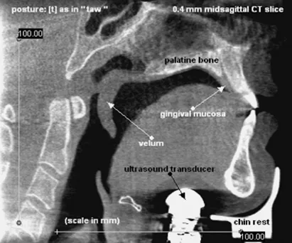
Computed Tomography uses X-rays to image slices (sections) of the body as thin as 0.5 mm or less. Tomographic images made in coronal planes are made by projecting very thin X-ray beams through a slice of tissue from multiple origins. The scanner rotates around the body taking these images and a computer creates a composite, including structures that are visible in some scans but obscured in others. Using this technique, tissue slices can be collected rapidly, 15 Hz or faster, and multiple slices can be collected simultaneously. CT images soft tissue more clearly than X-rays because it produces a composite X-ray. By digitally summing a series of scans, the composite section has sharper edges and more distinct tissue definition. From the multislice datasets, planar sections can be reconstructed in any direction. CT images can produce excellent resolution of soft and hard tissue structures. Figure 1.2, for example, is a reconstructed image of the midsagittal plane of the vocal tract. Bone appears bright white in the image, soft tissue structures are gray. In this figure, the junction of the velum and hard palate can be seen to be quite complex. The soft tissue below the hard palate widens before the velum emerges as a freestanding object. It is clear from this image that the shape of the palatine bone is not well reflected in the soft tissue. Measures of the palate bone made from an MRI or ultrasound image will differ from measurements made directly in the mouth or from dental impressions. Without this image, those differences would be hard to interpret.
Figure 1.2 Midsagittal CT of vocal tract reconstructed from axial images. Bone is white; soft tissue is gray.
(Reproduced courtesy of Ian Wilson)
Another method of CT data collection is Spiral CT. Spiral CT collects multiple slices at the same time by collecting a single spiral-shaped slice instead of multiple flat planar slices. In the mid 1980s, the cable and drum mechanism for powering the rotation of the CT machine was replaced with a slip ring. The slip ring allows the CT scanner to rotate continuously, creating a spiral image. Spiral CT scans have very high resolution, but currently take 20–30 seconds to create, and hence are too slow for imaging continuous speech, though excellent for st...
Table of contents
- Cover
- Praise for The Handbook of Phonetic Sciences
- Blackwell Handbooks in Linguistics
- Title page
- Copyright page
- Dedication
- Contributors
- Preface to the Second Edition
- Introduction
- Part I: Experimental Phonetics
- Part II: Biological Perspectives
- Part III: Modeling Speech Production and Perception
- Part IV: Linguistic Phonetics
- Part V: Speech Technology
- Index
Frequently asked questions
Yes, you can cancel anytime from the Subscription tab in your account settings on the Perlego website. Your subscription will stay active until the end of your current billing period. Learn how to cancel your subscription
No, books cannot be downloaded as external files, such as PDFs, for use outside of Perlego. However, you can download books within the Perlego app for offline reading on mobile or tablet. Learn how to download books offline
Perlego offers two plans: Essential and Complete
- Essential is ideal for learners and professionals who enjoy exploring a wide range of subjects. Access the Essential Library with 800,000+ trusted titles and best-sellers across business, personal growth, and the humanities. Includes unlimited reading time and Standard Read Aloud voice.
- Complete: Perfect for advanced learners and researchers needing full, unrestricted access. Unlock 1.5M+ books across hundreds of subjects, including academic and specialized titles. The Complete Plan also includes advanced features like Premium Read Aloud and Research Assistant.
We are an online textbook subscription service, where you can get access to an entire online library for less than the price of a single book per month. With over 1.5 million books across 990+ topics, we’ve got you covered! Learn about our mission
Look out for the read-aloud symbol on your next book to see if you can listen to it. The read-aloud tool reads text aloud for you, highlighting the text as it is being read. You can pause it, speed it up and slow it down. Learn more about Read Aloud
Yes! You can use the Perlego app on both iOS and Android devices to read anytime, anywhere — even offline. Perfect for commutes or when you’re on the go.
Please note we cannot support devices running on iOS 13 and Android 7 or earlier. Learn more about using the app
Please note we cannot support devices running on iOS 13 and Android 7 or earlier. Learn more about using the app
Yes, you can access The Handbook of Phonetic Sciences by William J. Hardcastle, John Laver, Fiona E. Gibbon, William J. Hardcastle,John Laver,Fiona E. Gibbon,John Laver in PDF and/or ePUB format, as well as other popular books in Languages & Linguistics & Phonetics & Phonology. We have over 1.5 million books available in our catalogue for you to explore.