![]()
1
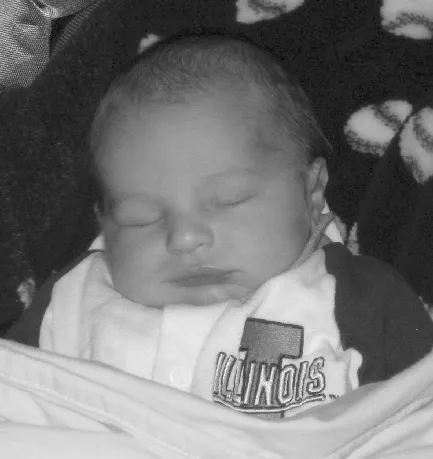
Figure 1.1 Matthew: Newborn
The Basic Biology of Brain Development
The brain is without doubt our most fascinating organ. Parents, educators, and society as a whole have a tremendous power to shape the wrinkly universe inside each child’s head, and, with it, the kind of person he or she will turn out to be. We owe it to our children to help them grow the best brains possible.
—L. Eliot (1999)
Brain Briefing
Although the brain is the least developed organ at birth, the baby has already started making connections to Mom through both smell and sound (Rodriguez, 2007).
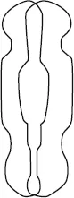
Brain development begins shortly after conception. Yes, Jack’s brain was busily creating itself from what is called the neural tube (Figure 1.2). This tube closed after about three weeks of gestation and proceeded to form itself into the miraculous structure we call the brain. Neurogenesis, the birth of neurons, proceeds rapidly. Since a baby is born with 100 billion neurons, they must be growing at a rate of over half a million per minute! (Eliot, 1999)
Figure 1.2 The Neural Tube That Will Form Into the Central Nervous System
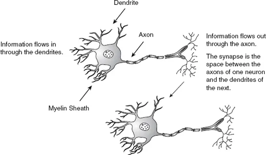
The neural tube is made up of cells that will give rise to the central nervous system. There are two different types of cells of which to be aware. Neurons are the brain cells that do most of the communicating in the brain and that we associate most with learning. Glial cells are support cells. They remove unneeded debris, and some literally wrap themselves around the output fiber of the neuron known as the axon. This white, sticky wrapper is called the myelin sheath. Myelin reinforces this message-sending appendage so that information moves faster and more securely. Besides having an axon that can send messages, neurons have dendrites to receive messages. Dendrites are the fibers that receive the information that has been sent out through another neuron’s axon. Remember: In through the dendrite; out through the axon! (Figure 1.3)
So, here we are with this teeny brain developing from the embryonic stage through the fetal stage. As the brain forms from the neural tube, neurons migrate to specific areas to learn to perform interesting tasks. For instance, some go to the occipital lobe in the back of the brain and become visual neurons. It is during this migration that the brain is highly susceptible to toxins such as alcohol. A pregnant woman drinking alcohol at crucial times can cause the neurons to change their migratory pattern. One result of this can be fetal alcohol syndrome.
Figure 1.3 Neuron
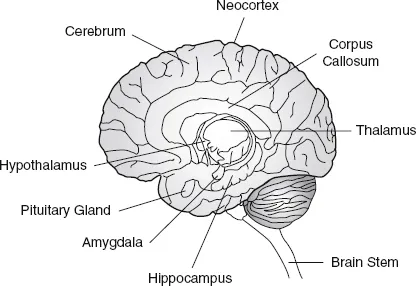
These brain cells migrate to become specific structures in the brain, from the brain stem that receives incoming sensory information (except for the sense of smell) all the way up to the neocortex that does our higher levels of thinking. The neocortex, also called the cerebral cortex, makes up about 80 percent of the brain’s volume (Figure 1.4). It is the outer layer of the brain that we sometimes call our gray matter.
Beneath the neocortex is a subcortical area called the limbic system. It consists of the thalamus, hypothalamus, hippocampus, and amygdala. The hypothalamus deals with internal communication between the body and the brain. The thalamus is a relay station for incoming information. It sends the messages to the appropriate places in the brain.
Figure 1.4 View of the Brain
In particular we will follow the growth of
• the hippocampus, which helps us form long-term factual memories.
• the amygdala, which filters all incoming information for emotional content.
• the corpus callosum, which connects the two hemispheres of the brain.
The brain is divided into a left and a right hemisphere. The hemispheres work together, yet each has some specific functions. Table 1.1 lists the information processing functions of each. Emotions and reading are just two areas that depend on the connections or “cross-talk” of these hemispheres (Elias & Arnold, 2006; Kagan & Herschkowitz, 2004).
Table 1.1 Functions of the Two Hemispheres Help Us Determine Specific Child Development Growth as the Two Hemispheres Grow and Connect
| Left Hemisphere | Right Hemisphere |
| Logical | Holistic |
| Details | Big picture |
| Language: speech, grammar, sounds | Language: prosody, tone |
| Expressive and receptive language | |
| Verbal short-term memory | Sensory image memory |
| Secondary processing of the | Secondary processing of emotional |
| expression of pleasurable emotion | communication: sending of unpleasurable |
| emotional signals; reception of both |
| pleasant and uncomfortable feelings |
| Reading body language |
| Facts | Events |
| Abstract processing | Concrete processing |
| Knowledge | Emotional significance of knowledge |
Each hemisphere is divided into lobes. The lobes have distinct functions. As we watch the growing brain develop, these lobes will be discussed. The development of each region has its own timetable. With the proper stimulation, the lobes mature to create a unique brain. Figure 1.5 shows the brain’s lobes, which include the following:
Occipital lobe. This lobe is responsible for vision. It is usually fully developed by age six.
Parietal lobe. It plays a part in the reception of sensory information. Also, the parietal lobe becomes active when problem solving and some calculations are attempted.
Brain Briefing
Touch activates many areas of the brain. Premature babies who were not touched and caressed did not develop as well or as fast as those who were
(Rodriguez, 2007).
Temporal lobe. This lobe is responsible for hearing, some speech, and some memories.
Frontal lobe. This is the area responsible for controlling emotions, working memory, decision making, future planning, verbal expression, and voluntary movement. The frontal lobe can be further divided. We often refer to the area behind the forehead, which is called the prefrontal cortex. It is here that emotion is modulated, feelings are managed, and attention is focused (Sunderland, 2006).
Figure 1.5 Lobes of the Brain
Also in Figure 1.5 you can see two important areas concerned with language and reading. Broca’s area in the frontal lobe is responsible for expressive language. It puts words together syntactically and grammatically. Wernicke’s area, in the parietal-temporal lobe, is responsible for receptive language. It stores the mental ...