![]()
Part I
Origin of Electrical Membrane Potential
This book is about the physiological characteristics of nerve and muscle cells. As we shall see, the ability of these cells to generate and conduct electricity is fundamental to their functioning. Thus, to understand the physiology of nerve and muscle, we must understand the basic physical and chemical principles underlying the electrical behavior of cells.
Because an understanding of how electrical voltages and currents arise in cells is central to our goals in this book, Part I is devoted to this task. The discussion begins with the differences in composition of the fluids inside and outside cells and culminates in a quantitative understanding of how ionic gradients across the cell membrane give rise to a transmembrane voltage. This quantitative description sets the stage for the specific descriptions of nerve and muscle cells in Parts II and III of the book and is central to understanding how the nervous system functions as a transmitter of electrical signals.
![]()
1
Introduction to Electrical Signaling in the Nervous System
The Patellar Reflex as a Model for Neural Function
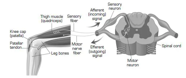
To set the stage for discussing the generation and transmission of signals in the nervous system, it will be useful to describe the characteristics of those signals using a simple example: the patellar reflex, also known as the knee-jerk reflex. Figure 1-1 shows the neural circuitry underlying the patellar reflex. Tapping the patellar tendon, which connects the knee cap (patella) to the bones of the lower leg, pulls the knee cap down and stretches the quadriceps muscle at the front of the thigh. Specialized nerve cells (sensory neurons) sense the stretch of the muscle and send a signal that travels along the thin fibers of the sensory neurons from the muscle to the spinal cord. In the spinal cord, the sensory signal is received by other neurons, called motor neurons. The motor neurons send nerve fibers back to the quadriceps muscle and command the muscle to contract, which causes the knee joint to extend.
Figure 1-1 A schematic representation of the patellar reflex. The sensory neuron is activated by stretching the thigh muscle. The incoming (afferent) signal is carried to the spinal cord along the nerve fiber of the sensory neuron. In the spinal cord, the sensory neuron activates motor neurons, which in turn send outgoing (efferent) signals along the nerve back to the thigh muscle, causing it to contract.
The reflex loop exemplified by the patellar reflex embodies in a particularly simple way all of the general features that characterize the operation of the nervous system. A sensory stimulus (muscle stretch) is detected, the signal is transmitted rapidly over long distance (to and from the spinal cord), and the information is focally and specifically directed to appropriate targets (the quadriceps motor neurons, in the case of the sensory neurons, and the quadriceps muscle cells, in the case of the motor neurons). The sensory pathway, which carries information into the nervous system, is called the afferent pathway, and the motor output constitutes the efferent pathway. Much of the nervous system is devoted to processing afferent sensory information and then making the proper connections with efferent pathways to ensure that an appropriate response occurs. In the case of the patellar reflex, the reflex loop ensures that passive stretch of the muscle will be automatically opposed by an active contraction, so that muscle length remains constant.
The Cellular Organization of Neurons
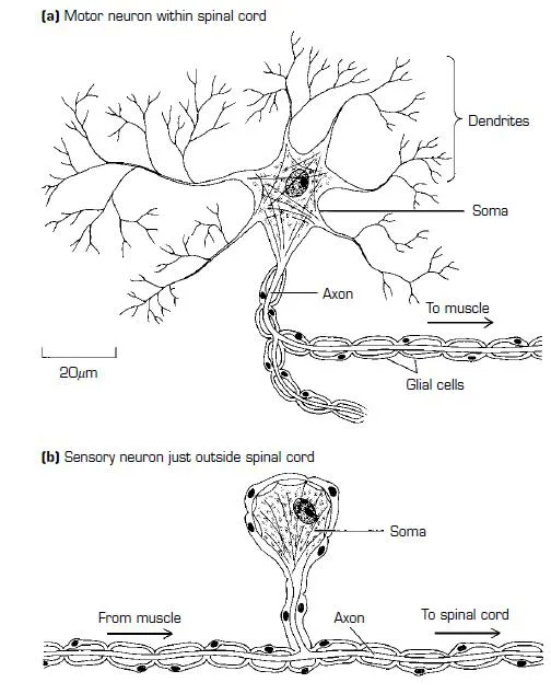
Neurons are structurally complex cells, with long fibrous extensions that are specialized to receive and transmit information. This complexity can be appreciated by examining the structure of a motor neuron, shown schematically in Figure 1-2a. The cell body, or soma, of the motor neuron—where the nucleus resides—is only about 20–30 µm in diameter in the case of motor neurons involved in the patellar reflex. The soma is only a small part of the neuron, however, and it gives rise to a tangle of profusely branching processes called dendrites, which can spread out for several millimeters within the spinal cord. The dendrites are specialized to receive signals passed along as the result of the activity of other neurons, such as the sensory neurons of the patellar reflex, and to funnel those signals to the soma. The soma also gives rise to a thin fiber, the axon, that is specialized to transmit signals over long distances. In the case of the motor neuron in the patellar reflex, the axon extends all the way from the spinal cord to the quadriceps muscle, a distance of approximately 1 meter. As shown in Figure 1-2b, the sensory neuron of the patellar reflex is structurally simpler than the motor neuron. Its soma, which is located just outside the spinal cord in the dorsal root ganglion, gives rise to only a single nerve fiber, the axon. The axon splits into two branches shortly after it exits the dorsal root ganglion: one branch extends away from the spinal cord to contact the muscle cells of the quadriceps muscle, and the other branch passes into the spinal cord to contact the quadriceps motor neurons. The axon of the sensory neuron carries the signal generated by muscle stretch from the muscle into the spinal cord. Because the sensory neuron receives its input signal from the sensory stimulus (muscle stretch) at the peripheral end of the axon instead of from other neurons, it lacks the den-drites seen in the motor neuron.
Figure 1-2 Structures of single neurons involved in the patellar reflex.
Electrical Signals in Neurons
To transmit information rapidly over long distances, neurons produce active electrical signals, which travel along the axons that make up the transmission paths. The electrical signal arises from changes in the electrical voltage difference across the cell membrane, which is called the membrane potential.Although this transmembrane voltage is small—typically less than a tenth of a volt—it is central to the functioning of the nervous system. Information is transmitted and processed by neurons by means of changes in the membrane potential.
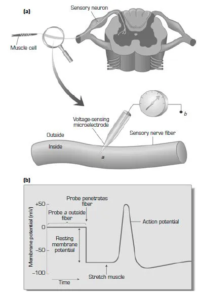
What does the electrical signal that carries the message along the sensory nerve fiber in the patellar reflex look like? To answer this question, we must measure the membrane potential of the sensory neuron by placing an ultrafine voltage-sensing probe, called an intracellular microelectrode, inside the sensory nerve fiber, as illustrated in Figure 1-3. A voltmeter is connected to measure the voltage difference between the tip of the intracellular microelectrode (point a in the figure) and a reference point in the extracellular space (point b). When the microelectrode is located outside the sensory neuron, both points a and b are in the extracellular space, and the voltmeter therefore records no voltage difference (Figure 1-3b). When the tip of the probe is inserted inside the sensory neuron, however, the voltmeter measures an electrical potential between points a and b, representing the voltage difference between the inside and the outside of the neuron—that is, the membrane potential of the neuron. As shown in Figure 1-3b, the inside of the sensory nerve fiber is negative with respect to the outside by about seventy-thousandths of a volt (1 millivolt, abbreviated mV, equals one-thousandth of a volt). Because the potential outside the cell is our reference point and the inside is negative with respect to the outside, the membrane potential is represented as a negative number, i.e., –70 mV.
As long as the sensory neuron is not stimulated by stretching the muscle, the membrane potential remains constant at this resting value. For this reason, the unstimulated membrane potential is known as the resting potential of the cell. When the muscle is stretched, however, the membrane potential of the sensory neuron undergoes a dramatic change, as shown in Figure 1-3b. After a delay that depends on the distance of the recording site from the muscle, the membrane potential suddenly moves in the positive direction, transiently reverses sign for a brief period, and then returns to the resting negative level. This transient jump in membrane potential is the action potential—the long-distance signal that carries information in the nervous system.
Transmission between Neurons
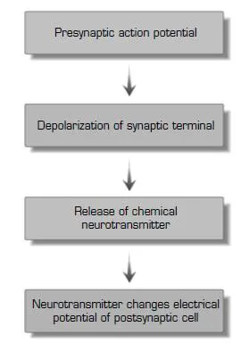
What happens when the action potential reaches the end of the neuron, and the signal must be transmitted to the next cell? In the patellar reflex, signals are relayed from one cell to another at two locations: from the sensory neuron to the motor neuron in the spinal cord, and from the motor neuron to the muscle cells in the quadriceps muscle. The point of contact where signals are transmitted from one neuron to another is called a synapse. In the patellar reflex, both the synapse between the sensory neuron and the motor neuron and the synapse between the motor neuron and the muscle cells are chemical synapses, in which an action potential in the input cell (the presynaptic cell) causes it to release a chemical substance, called a neurotransmitter. The molecules of neuro-transmitter then diffuse through the extracellular space and change the membrane potential of the target cell (the postsynaptic cell). The change in membrane potential of the target then affects the firing of action potentials Neurotransmitter changes electrica potential of postsynaptic cell by the postsynaptic cell. This sequence of events during synaptic transmission is summarized in Figure 1-4.
Figure 1-3 Recording the action potential in the nerve fiber of the sensory neuron in the patellar stretch reflex. (a) A diagram of the recording configuration. A tiny microelectrode is inserted into the sensory nerve fiber, and a voltmeter is connected to measure the voltage difference (E) between the inside (a) and the outside (b) of the nerve fiber. (b) When the microelectrode penetrates the fiber, the resting membrane potential of the nerve fiber is measured. When the sensory neuron is activated by stretching the muscle, an action potential occurs and is recorded as a rapid shift in the recorded membrane potential of the sensory nerve fiber.
Figure 1-4 Chemical transmission mediates synaptic communication between cells in the patellar reflex. The flow diagram shows the sequence of events involved in the release of chemical neurotransmitter from the synaptic terminal.
Because signaling both within and between cells in the nervous system involves changes in the membrane potential, the brain is essentially an electrochemical organ. Therefore, to understand how the brain functions, we must first understand the electrochemical mechanisms that give rise to a transmembrane voltage in cells. The remaining chapters in Part I are devoted to the task of developing the basic chemical and physical principles required to comprehend how cells communicate in the nervous system. In Part II, we will then consider how these electrochemical principles are exploited in the nervous system for both long-distance communication via action potentials and local communication at synapses.
![]()
2
Composition of Intracellular and Extracellular Fluids
When we think of biological molecules, we normally think of all the special molecules that are unique to living organisms, such as proteins and nucleic acids: enzymes, DNA, RNA, and so on. These are the substances that allow life to occur and that give living things their special characteristics. Yet, if we were to dissociate a human body into its component molecules and sort them by type, we would find that these special molecules are only a small minority of the total. Of all the molecules in a human body, only about 0.25% fall within the category of these special biological molecules. Most of the molecules are far more ordinary. In fact, the most common molecule in the body is water. Excluding nonessential body fat, water makes up about 75% of the weight of a human body. Because water is a comparatively light molecule, especially when compared with massive protein molecules, this 75% of body weight translates into a staggering number of molecules of water. Thus, water molecules account for about 99% of all molecules in the body. The remaining 0.75% consists of other simple inorganic substances, mostly sodium, potassium, and chloride ions. In the first part of this book we will be concerned in large part with the mundane majority of molecules, the 99.75% made up of water and inorganic ions.
Why should we study these mundane molecules? Many enzymatic reactions involving the more glamorous organic molecules require the participation of inorganic cofactors, and most biochemical reactions within cells occur among substances that are dissolved in water. Nevertheless, most inorganic molecules in the body never participate in any biochemical reactions. In spite of this, a sufficient reason to study these inorganic substances is that cells could not exist and life as we know it would not be possible if cells did not possess mechanisms to control the distribution of water and ions across their membranes. The purpose of this chapter is to see why that is true and to understand the physical principles that unde...