![]()
1
The Digestive System
A horse which is kept to dry meat will often slaver at the mouth. If he champs his hay and corn, and puts it out again, it arises from some fault in the grinders… there will sometimes be great holes cut with his grinders in the weaks of his mouth. First file his grinders quite smooth with a file made for the purpose.
Francis Clater, 1786
Horses are ungulates and, according to J.Z. Young (1950), members of the order Perissodactyla. Other extant members include asses, zebras, rhinoceroses and tapirs. Distinctive characteristics of the order are the development of the teeth, the lower limb with the peculiar plan of the carpus and tarsus bones and the evolution of the hind gut into chambers for fermentation of ingesta. Each of these distinctive features will play significant roles in the discussions in this text.
The domesticated horse consumes a variety of feeds, ranging in physical form from forage with a high content of moisture to cereals with large amounts of starch, and from hay in the form of physically long fibrous stems to salt licks and water. In contrast, the wild horse has evolved and adapted to a grazing and browsing existence, in which it selects succulent forages containing relatively large amounts of water, soluble proteins, lipids, sugars and structural carbohydrates, but little starch. Short periods of feeding occur throughout most of the day and night, although generally these are of greater intensity in daylight. In domesticating the horse, man has generally restricted its feeding time and introduced unfamiliar materials, particularly starchy cereals, protein concentrates and dried forages. The art of feeding gained by long experience is to ensure that these materials meet the varied requirements of horses without causing digestive and metabolic upsets. Thus, an understanding of the form and function of the alimentary canal is fundamental to a discussion of feeding and nutrition of the horse.
THE MOUTH
Eating rates of horses, cattle and sheep
The lips, tongue and teeth of the horse are ideally suited for the prehension, ingestion and alteration of the physical form of feed to that suitable for propulsion through the gastrointestinal (GI) tract in a state that facilitates admixture with digestive juices. The upper lip is strong, mobile and sensitive and is used during grazing to place forage between the teeth; in the cow the tongue is used for this purpose. By contrast, the horse’s tongue moves ingested material to the cheek teeth for grinding. The lips are also used as a funnel through which water is sucked.
As distinct from cattle, the horse has both upper and lower incisors enabling it to graze closely by shearing off forage. More intensive mastication by the horse means that the ingestion rate of long hay, per kg of metabolic body weight (BW), is three to four times as fast in cattle and sheep than it is in ponies and horses, although the number of chews per minute is similar, according to published observations (73–92 for horses and 73–115 for sheep) for long hays. The dry matter (DM) intake per kg of metabolic BW for each chew is then 2.5 mg in horses (I calculate it to be even less – author) and 5.6–6.9mg in sheep. Consequently, the horse needs longer daily periods of grazing than do sheep. The lateral and vertical movements of the horse’s jaw, accompanied by profuse salivation, enable the cheek teeth to comminute long hay to a large extent and the small particles coated with mucus are suitable for swallowing. Sound teeth generally reduce hay and grass particles to less than 1.6 mm in length. Two-thirds of hay particles in the horse’s stomach are less than 1 mm across, according to work by Meyer and colleagues (Meyer et al. 1975b).
The number of chewing movements for roughage is considerably greater than that required for chewing concentrates. Horses make between 800 and 1200 chewing movements per 1 kg concentrates, whereas 1 kg long hay requires between 3000 and 3500 movements. In ponies, chewing is even more protracted – they require 5000–8000 chewing movements per 1 kg concentrates alone, and very many more for hay (Meyer et al. 1975b). Horses given a hay diet chewed 40,000 times/day compared with 10,000 times/day for those fed on pellets (Houpt et al. 2004). Hay chewing, cf. pellets, by both horses and ponies, is protracted, with a lower chewing-cycle frequency, as the mandibular displacement is greater, both vertically and horizontally with an effect on faecal particle dimensions (Brøkner et al. 2009). Clayton et al. (2003) concluded that the development of sharp enamel points is more likely with a high concentrate diet.
Mature and young horses have a maximal daily DM intake of 3.0–3.2% of BW, although the average is lower (NRC 2007). Ponies have a higher voluntary DM intake than horses; Pearson et al. (2001) found ponies ate 3.9 kg/100 kg BW alfalfa hay while Argo et al. (2002) recorded 5.1 kg fresh weight/100 kg BW of a meal of 60% hay and 40% concentrate pellets. Such high intakes might occur with high quality feed after a period of feed restriction, as particle retention time is greater for poor quality feed (Pearson et al. 2001). The addition of 35% short chaff (<2 cm) to sweet coarse mix slowed the rate of consumption and doubled the eating time, but increased the eating rate (Harris et al. 2005) and the addition of chopped straw, either 2.5 or 4 cm in length at rates of 10–30% of a pelleted diet mixed with chopped alfalfa, increased the time to eat 1 kg wet matter (Ellis et al. 2005). These observations are important for an understanding of healthy digestion.
Dentition
As indicated above, teeth are vital to the well-being of horses. Diseased teeth are an encumbrance. Primary disorders of the cheek teeth represented 87% of the dental disorders in 400 horses (Dixon et al. 2000a). The disorders included abnormalities of wear, traumatic damage, and fractures from which the response to treatment was good. Dental and head pain have specific behavioural indicators, including altered eating patterns, anorexia, feed refusal and quidding (Ashley et al. 2005) and cause digestive disturbances and colic. The prevalence of dental disorders amongst donkeys increases with age, and is especially prominent at 15–20 years of age. Dental disease is associated with poor body condition score (BCS), previous episodes of colic, diastemata (a gap between adjacent teeth) and wave-, smooth- and step-mouth (Du Toit et al 2009a,b).
Apparent fibre digestibility, the proportion of faecal short fibre particles and plasma free fatty acids (FFAs) were all increased after dental correction in mares. Consequently, diseased and badly worn teeth, as in the geriatric horse, can limit the horse’s ability to handle roughage, that compromises general health. Infections of cheek teeth are not uncommon and Dixon et al. (2000b) found that nasal discharge was more frequent with infections of caudal than with rostral maxillary teeth. Hudson et al (2006) describe cases of dysphagia in horses caused by a buccal abscess, a lingual abscess, a retropharyngeal foreign body and an oesophageal obstruction. Windley et al (2009b) reported that both twoand threedimensional computed tomography (CT) were valuable as clinical diagnostic tools in detection of dental lesions and in selection of appropriate treatment.
The apparent digestibility of the protein and fibre in hay and grain is reduced if the occlusal angle of premolar 307 is greater than 80° relative to the (flattened) vertical angle (Ralston et al. 2001). However, no adverse effects were noted by Carmalt & Allen (2008) where normal variation occurs in occlusial characteristics; they found no relationships between cheek tooth occlusal morphology, apparent feed digestibility, and the reduction in particle size of three different hay-based feeds.
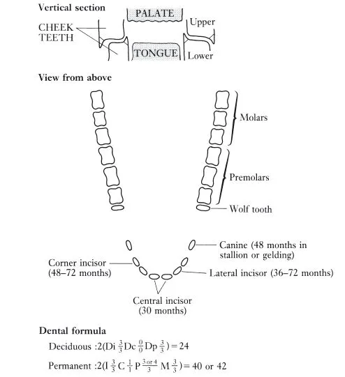
The normal horse has two sets of teeth. The first to appear, the deciduous, or temporary milk, teeth erupt soon after birth and are replaced during growth by the permanent teeth. The permanent incisors and cheek teeth erupt continuously to compensate for wear, and their changing form provides a basis for assessing the age of a horse. In the gap along the jaw between the incisors and the cheek teeth the male horse normally has a set of small canine teeth. The gap, by happy chance, securely locates the bit. The dental formulae and configuration of both deciduous and permanent teeth are given in Figure 1.1. The lower cheek teeth are implanted in the mandible in two straight rows that diverge towards the back. The space between the rows of teeth in the lower jaw is less than that separating the upper teeth (Figure 1.1). This accommodates a sideways, or circular, movement of the jaw that effectively shears feed. The action leads to a distinctive pattern of wear of the biting surface of the exposed crown. This pattern results from the differences in hardness which characterize the three materials (cement, enamel and dentine) of which teeth are composed. The enamel, being the hardest, stands out in the form of sharp prominent ridges. It is estimated that the enamel ridges of an upper cheek tooth in a young adult horse, if straightened out, would form a line more than 30 cm (1 ft) long. This irregular surface provides a very efficient grinding organ.
Figure 1.1 Configuration of permanent teeth in the upper or lower jaw (the molars and premolars in the lower jaw are slightly closer to the midline). The deciduous teeth on each side of each jaw consist of three incisors, one canine, and three molars. The deciduous canines are vestigial and do not erupt. The wolf teeth (present in the upper jaw of about 30% of fillies and about 65% of colts) are often extracted, as their sharp tips can injure cheeks when a snaffle bit is used. Months (in parentheses) are approximate ages at which permanent incisors and canines erupt, replacing the deciduous teeth.
Horses and ponies rely more on their teeth than we do. The human diet could be said to consist mainly of concentrates, which require much less chewing than does roughage. A dietary regime consisting mainly of concentrate feeds is associated with smaller mandibular excursions during chewing by the horse. This could imply that, during training, more frequent dental prophylactic treatment is needed to avoid development of dental irregularities (Bonin et al. 2007). Even among herbivores, horses and ponies depend to a far greater extent on their teeth than do the domesticated ruminants – cattle, sheep and goats. Ruminants, as discussed in ‘Eating rates of horses, cattle and sheep. above, swallow grass and hay with minimal chewing and then depend on the activity of bacteria in the rumen to disrupt the fibre. The fibre is then much more readily fragmented during chewing the cud.
Saliva
The physical presence of feed material in the mouth stimulates the secretion of a copious amount of saliva. Some 10–12L are secreted daily in a normally fed horse. This fluid seems to have no digestive enzyme activity, but its mucus content enables it to function as an efficient lubricant preventing ‘choke’. Its bicarbonate content, amounting to some 50 mEq/L, provides it with a buffering capacity. The production of bicarbonate and sodium chloride in the saliva is directly proportional to the rate of secretion. The continuous secretion of saliva during eating seems to buffer the digesta in the proximal region of the stomach, permitting some microbial fermentation with the production of lactate. This has important implications for the well-being of the horse (see Chapter 11).
Obstruction of the oesophagus by impacted feed or foreign bodies is not uncommon, but attempts to pass a nasogastric tube are not justified, as most cases respond to conservative treatment. For cases of more than 48 h duration a cuffed nasogastric tube is advocated, although the value of oxytocin use is unclear (Duncanson 2006.. To facilitate nutritional support during treatment of oesophageal perforation, a cervical oesophagotomy tube is placed and advanced into the stomach (Read et al. 2002). An enteral diet includes an electrolyte mixture (partly to compensate for salivary electrolyte losses through the oesophagotomy site), sucrose (1.2 kg/day), casein, canola rapeseed oil (1.1L/day) and dehydrated alfalfa pellets. A nasogastric tube is subsequently introduced to allow repair of the oesophagotomy site.
THE STOMACH AND SMALL INTESTINE
The first quantitative aspects of digestion were demonstrated by Waldinger in 1808 with the passage of capsulated feedstuff through the intestines. Intensive studies concerning the physiology of digestion were started in Paris around 1850 by Colin, but they were continued predominately from 1880 in Dresden by Ellenberger and Hofmeister who investigated the mouth, stomach and small intestine. Scheunert continued with work on the large intestine in Dresden and Leipzig until the 1920s. Although the apparent digestibility of cellulose was appreciated by 1865 it took another 20 years for the discovery of the process of microbial digestion in the equine large intestine. Until 1950 most routine equine digestibility experiments were conducted in Germany, France and the USA (Klingeberg-Kraus 2001), while comparative studies were conducted by Phillipson, Elsden and colleagues at Cambridge in the 1940s.
Development of the gastrointestinal (GI) tract and associated organs
The GI tract tissue of the neonatal foal weighs only 35 g/kg BW, whereas the liver is large, nearly in the same proportion to BW, acting as a nutrient store for the early critical days. By six months of age the GI tract tissue has proportionately increased to 60 g/kg BW, whereas the liver has proportionately decreased to about 12–14 g/kg BW. By 12 months both these organs have stabilized at 45–50 g/kg BW for the GI tract and 10 g/kg BW for the liver. Organ size is also influenced by the activity of the horse. After a meal, the liver of mammals generally increases rapidly in weight, probably as a result of glycogen storage and blood flow. In the horse the consumption of hay has less impact on liver glycogen, so that following a meal of hay the liver weighs only three-quarters of that following mixed feed. Moreover, during and immediately after exercise the GI tract tissue weighs significantly less than in horses at rest, owing to the shunting of blood away from the mesenteric blood vessels to the muscles. At rest, about 30% of the cardiac output flows through the hepatic portal system. These aspects are discussed further in Chapter 9.
Surprisingly, the small intestine does not materially increase in length from 4 weeks of age, whereas the large intestine increases with age, the colon doing so until 20 years at least. The distal regions of the large intestine continue extension to a greater age than do the proximal regions. This development reflects the increasing reliance of the older animal on roughage. In an adult horse of 500 kg BW the small intestine is approximately 16 m in length, the caecum has a maximum length of about 0.8 m, the ascending colon 3 m and the descending colon 2.8 m.
Transit of digesta through the GI tract
The residence time for ingesta in each section of the GI tract allows for its adequate admixture with GI secretions, for hydrolysis by digestive enzymes, for absorption of the resulting products, for fermentation of resistant material by bacteria and for the absorption of the products of that fermentation. Transit time through the GI tract is normally considered in three phases, owing to their entirely different characteristics. These phases are:
(1) expulsion rate from the stomach into the duodenum after a meal;
(2) rate of passage through the small intestine to the ileocaecal orifice;
(3) retention time in the large intestine.
The first of these will be considered below in relation to gastric disorders. Rate of passage of digesta through the small intestine varies with feed type. On pasture this rate is accelerated, although a previous feed of hay causes a decrease in the rate of the succeeding meal, with implications for exercise (see Chapter 9). Roughage is held in the large intestine for a considerable period that allows microbial fermentation time to break down structural carbohydrates. However, equine GI transit time of the residue of high fibre diets is less than that of low fibre diets of the same particle size, in common with the relationship found in other monogastric animals.
Digestive function of the stomach
The stomach of the adult horse is a small organ, its volume comprising about 10% of the GI tract (Figure 1.2, Plate 1.1; Meyer et al. 1993a). In the suckling foal, however, the stomach capacity represents a larger proportion of the total alimentary tract. Most digesta are held in the stomach for a comparatively short time, but this organ is rarely completely empty and a significant portion of the digesta remain in it for 2 to 6 h. Some digesta pass into the duodenum shortly after eating starts, when fresh ingesta enter the stomach. Expulsion into the duodenum is arrested as soon as feeding stops. When a horse drinks, a high proportion of the water passes along the curvature of the stomach wall so that mixing with digesta and dilution of the digestive juices it contains are avoided. This process is particularly noticeable when digesta fill the stomach.
The entrance to the stomach is guarded by a powerful muscular valve called the cardiac sphincter. Although a horse might feel nauseated, it rarely vomits, partly because of the way this valve...