Pocket Guide to Echocardiography
- English
- ePUB (mobile friendly)
- Available on iOS & Android
About this book
With its easy accessibility, low cost, and ability to deliver, essential bedside information about the cardiac structure and function, echocardiography has become one of the most relied-upon diagnostic tools in clinical medicine. As a result, more clinicians than ever before must be able to accurately interpret echocardiographic information in order to administer appropriate treatment.
Based on the authors' experience teaching echocardiography in busy clinical settings, this new pocketbook provides reliable guidance on everyday clinical cardiac ultrasound and the interpretation of echocardiographic images. It has been designed to help readers develop a stepwise approach to the interpretation of a standard transthoracic echocardiographic study and teach how to methodically gather and assemble the most important information from each of the standard echocardiographic views in order to generate a complete final report of the study performed.
What's included:
• A summary of TTE examination protocol and a comprehensive listing of useful formulas and normal values
• Atrial and ventricular dimensions, LV and RV systolic function, LV diastolic patterns
• Echocardiographic findings in the most commonly encountered cardiac diseases and disorders, including various cardiomyopathies, cardiac tamponade, constrictive pericarditis, valvular heart disease, pulmonary hypertension, infective endocarditis, and congenital heart disease
• Companion website with video clips and over 70 self-assessment questions
Packed with essential information and designed for quick look-up, this pocketbook will be of great assistance for anyone who works in busy clinical settings and who needs a ready and reliable guide to interpreting echocardiographic information to help deliver optimal patient care.
Frequently asked questions
- Essential is ideal for learners and professionals who enjoy exploring a wide range of subjects. Access the Essential Library with 800,000+ trusted titles and best-sellers across business, personal growth, and the humanities. Includes unlimited reading time and Standard Read Aloud voice.
- Complete: Perfect for advanced learners and researchers needing full, unrestricted access. Unlock 1.4M+ books across hundreds of subjects, including academic and specialized titles. The Complete Plan also includes advanced features like Premium Read Aloud and Research Assistant.
Please note we cannot support devices running on iOS 13 and Android 7 or earlier. Learn more about using the app.
Information
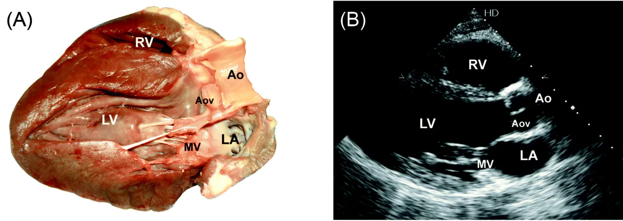
Parasternal Long-axis view (Fig. 1)
RA/RV view
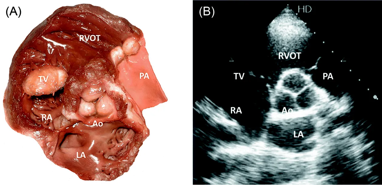
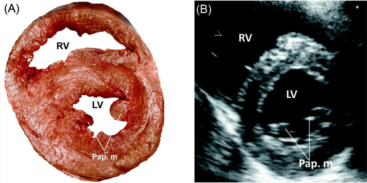
Parasternal Short-axis view (Fig. 2 and Fig. 3)
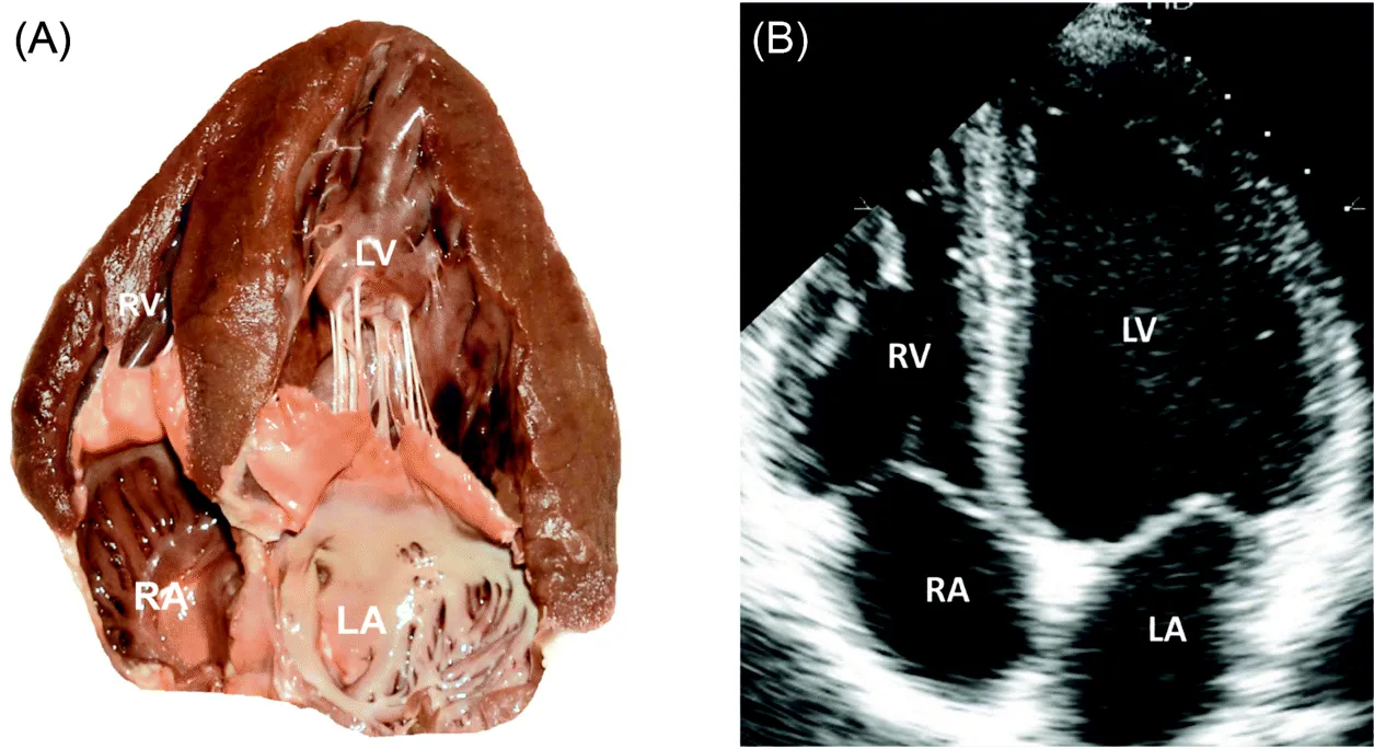
Apical 4-chambers view (Fig. 4)
Apical 5-chambers view
Table of contents
- Cover
- Companion Website
- Title Page
- Copyright
- Foreword by Navin C. Nanda
- Preface
- Abbreviations
- Chapter 1: Comprehensive Transthoracic Echocardiographic Examination Protocol
- Chapter 2: Indications, Contraindications and Endpoints of Dobutamine and Exercise Stress Echocardiography
- Chapter 3: Types of Stress Echocardiography and Reading Template
- Chapter 4: Useful Formulas and Normal Values
- Chapter 5: Guidelines for the Safe use of Echocardiography Contrast
- Chapter 6: Atrial and Ventricular Dimensions
- Chapter 7: Coronary Artery Disease
- Chapter 8: Left Ventricular Systolic Function and left Ventricular Diastolic Patterns
- Chapter 9: Right Ventricular Systolic Function and Right Ventricular Diastolic Patterns
- Chapter 10: Dilated, Hypertrophic and Restrictive Cardiomyopathies
- Chapter 11: Pericardial Effusion, Cardiac Tamponade, Constrictive Pericarditis
- Chapter 12: Mitral Stenosis
- Chapter 13: Mitral Valvuloplasty Score
- Chapter 14: Recommendations for data Recording and Measurement for Mitral Stenosis
- Chapter 15: Mitral Regurgitation
- Chapter 16: Aortic Regurgitation
- Chapter 17: Aortic Stenosis
- Chapter 18: Recommendations for Data Recording and Measurement for Aortic Stenosis
- Chapter 19: Resolution of Apparent Discrepancies in Measures of Aortic Stenois Severity
- Chapter 20: Pulmonic Stenosis, Pulmonic Regurgitation, Pulmonary Hypertension
- Chapter 21: Tricuspid Regurgitation and Tricuspid Stenosis
- Chapter 22: Infective Endocarditis
- Chapter 23: ACC/ASE Recommendations for Echocardiography in Ineffective Endocarditis
- Chapter 24: Prosthetic Valves
- Chapter 25: Normal Echocardiographic Values for Prosthetic Valves
- Chapter 26: Congenital Heart Disease
- Chapter 27: Miscellaneous
- Chapter 28: Aortic Diseases
- Chapter 29: Indication for Surgery in Aortic Diseases
- Chapter 30: Transthoracic Echocardiographic and Doppler Protocols for Assessment of Ventricular Dyssynchrony
- Chapter 31: Indications, Contraindications and Complications of Transesophageal Echocardiographic Examination
- Chapter 32: Routine Approach to any Transesophageal Echocardiographic and Recommended views for Evaluation of Aorta
- Chapter 33: Terminology used to Describe Manipulation of the Probe and Transducer During Image Acquisition
- Chapter 34: Diagrams of Standard Transesophageal Echocardiographic Views
- Chapter 35: Transesophageal Echocardiographic Measurements
- Chapter 36: Transesophageal Echocardiographic Diagram of the Regional Blood Supply to Cardiac Wall Segments
- Chapter 37: Transesophageal Echocardiographic Orientation for Assessment of the Mitral Valve
- Chapter 38: Diagrams of Transesophageal Echocardiographic views in the Evaluation of the Mitral Valve
- Chapter 39: References and Recommended Literature
- Supplement to Pocket Guide of Echocardiography