![]()
Part 1
Fundamentals
![]()
1
Introduction
Clinical background
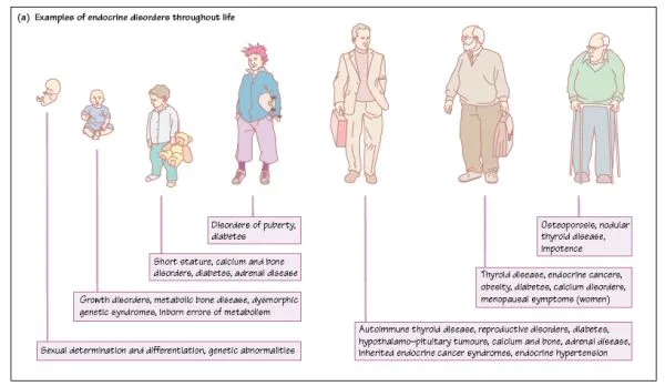
Endocrinology is the study of endocrine hormones and of the organs involved in endocrine hormone release. Classically, hormones have been described as chemical messengers, released and having their actions at distant sites. It is now clear, however, that there is a close relationship between hormones and other factors such as neurotransmitters and growth factors acting in a paracrine or autocrine fashion. Hormones are essential for the maintenance of normal physiological function and hormonal disorders occur at all stages of human life. Clinical endocrinologists thus look after patients of all ages and with a very wide range of disorders (Fig. 1a).
The principal endocrine glands
The brain is the controller of the nervous system, but it is also one of the most important endocrine glands. Specialized nerve cells, notably in the hypothalamus, synthesize hormones which are transported along the axon to the nerve terminal. Here they are released into the portal blood system, which carries them to the pituitary gland. In some cases, the axon of the neuroendocrine cell projects down to the pituitary cell itself. The principal hypothalamic neurohormones are:
1. corticotrophin-releasing hormone (CRH), controls the release of ACTH;
2. dopamine inhibits prolactin release;
3. growth-hormone-releasing hormone (GHRH) causes growth hormone release;
4. somatostatin inhibits growth hormone release;
5. gonadotrophin-releasing hormone (GnRH) causes luteinizing hormone (LH) and follicle- stimulating hormone (FSH) release;
6. thyrotrophin-releasing hormone (TRH) causes thyroid- stimulating hormone (TSH) release;
7. oxytocin causes milk ejection and contraction of the uterus in labour - it is synthesized in the hypothalamus and is stored in and released from the posterior pituitary gland;
8. vasopressin (antidiuretic hormone, ADH) promotes water reabsorption from the kidney tubules - it is synthesized in the hypothalamus, and stored in and released from the posterior pituitary gland.
The pituitary gland is composed of two lobes, anterior and posterior, which arise from different embryological origins - the anterior originates from the embryonic oral cavity and the posterior from the base of the brain (i.e. a neural origin). The two lobes become closely apposed to each other to form the pituitary gland. Humans have a non-functional intermediate lobe, which is much larger in some other animals. The principal hormones of the pituitary are:
1 anterior:
(a) corticotrophin (adrenocorticotrophic hormone; ACTH) releases glucocorticoids and other steroids from the adrenal cortex;
(b) follicle-stimulating hormone (FSH) promotes spermatogenesis in males and ovarian follicular maturation in females;
(c) luteinizing hormone (LH) promotes testosterone synthesis in males and causes ovarian follicular rupture and ovulation in females;
(d) prolactin (PRL) promotes lactation and may have an immunomodulatory role in non-lactating females and males;
(e) thyrotrophin (thyroid-stimulating hormone; TSH) promotes thyroid hormone production and release from the thyroid gland;
(f) growth hormone (also called somatotrophin; GH) promotes muscle and skeletal growth.
2 posterior:
(a) oxytocin causes milk ejection and contraction of the uterus in labour;
(b) vasopressin (antidiuretic hormone, ADH) promotes water reabsorption from the renal tubules.
The thyroid gland is situated just in front of the trachea in humans. The thyroid-hormone-producing cells are arranged in follicles, and concentrate iodine which is used for the synthesis of the thyroid hormone. The circulating hormones are thyrox- ine (T4) and tri-iodothyronine (T3). The parathyroid glands are embedded in the thyroid, and produce parathyroid hormone (parathormone; PTH). PTH is important in the control of calcium and phosphate metabolism. The parafollicular cells are in the thyroid, scattered between the follicles. They produce calcitonin, which inhibits bone calcium resorption.
The adrenal glands are situated just above the kidneys, and are composed of an outer layer, or cortex, and an inner layer, or medulla (a modified ganglion). The hormones produced are:
1 cortex:
(a) glucocorticoids, principally cortisol in humans, are involved in carbohydrate metabolism and the response to stress;
(b) mineralocorticoids, principally aldosterone, control electrolyte balance;
(c) androgens, principally testosterone, dehydroepiandros- tenedione sulphate (DHEAS) and 17-hydroxyprogesterone, modulate secondary sexual characteristics and have anabolic effects.
2 medulla:
(a) epinephrine modulates cardiovascular and metabolic response to stress;
(b) norepinephrine, principally a neurotransmitter in the peripheral sympathetic nervous system;
(c) dopamine, a neurotransmitter in the autonomic nervous system.
The endocrine pancreas consists of islet cells scattered in the larger exocrine pancreas, which lies posteriorly in the upper abdomen. (‘Exocrine’ refers to glands which have ducts, and which are not covered in this book.) The endocrine pancreas secretes:
1 insulin, which regulates glucose and lipid metabolism;
2 glucagon, a counter-regulatory hormone to insulin that elevates blood glucose;
3 somatostatin, which regulates gastrointestinal motility;
4 pancreatic polypeptide, which regulates gastrointestinal secretion.
The ovary is the major female reproductive gland, and produces:
1 estrogens, which regulate reproductive function and secondary sexual characteristics;
2 progesterone, which stimulates endometrial vascularization and maintains pregnancy;
3 relaxin, a polypeptide also found in the placenta and uterus, which may be important in parturition by softening the cervix and relaxing the pelvic ligaments;
4 inhibin, which inhibits FSH production.
The placenta is the organ of pregnancy serving the developing fetus. Hormones produced by the placenta include:
1 chorionic gonadotrophin (CG; hCG; h = human) which maintains placental progesterone synthesis;
2 placental lactogen (PL);
3 estriol, the major form of estrogen secreted by the placenta;
4 progesterone which maintains the reproductive organs in pregnancy;
5 relaxin.
The testis is the major male reproductive gland, producing:
1 testosterone which controls reproductive function and secondary sexual characteristics;
2 inhibin, which inhibits FSH secretion;
3 Mullerian inhibiting hormone (MIH), a fetal hormone which dedifferentiates the Mullerian duct.
The gastrointestinal tract (GIT) is the largest endocrine organ and produces several autocrine, paracrine and endocrine hormones including:
1 cholecystokinin (CCK);
2 gastric inhibitory peptide (GIP);
3 gastrin;
4 neurotensin;
5 secretin;
6 substance P;
7 vasoactive intestinal peptide (VIP).
Adipocytes produce the peptide hormone leptin which is important in the control of feeding and energy expenditure.
The kidney produces hormones involved in the control of blood pressure and in erythropoiesis. Renin cleaves angiotensinogen to angiotensin I in the kidney and plasma. Erythropoietin stimulates production of red blood cells in the marrow.
The skin, liver and kidney produce vitamin D which has certain endocrine functions.
The heart produces atrial natriuretic peptide. Circulating blood elements, including macrophages, produce peptides such as the cytokines, which are involved in immune function.
The pineal gland is situated in the brain and is involved with rhythms, for example the reproductive rhythms of animals which breed seasonally. Its role in humans is not known for certain. The pineal gland produces melatonin.
Readers should be aware that putative endocrine hormones continue to be reported.
![]()
2
Chemical transmission
Classification of endocrine hormones
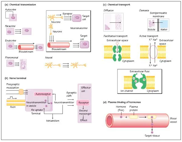
Hormones are chemical messengers. They may be classified several ways (Fig. 2a):
1 Autocrine: acting on the cells that synthesized them; for example insulin- like growth factor (IGF- 1) which stimulates cell division in the cell which produced it.
2 Paracrine: acting on neighbouring cells. An example is insulin, secreted by pancreatic b cells and affecting secretion of glucagon by pancreatic a cells.
3 Endocrine: acting on cells or organs to which they are carried in the bloodstream or through another aqueous ducting system, such as lymph. Examples include insulin, estradiol and cortisol.
4 Neuroendocrine: this is really paracrine or endocrine, except that the hormones are synthesized in a nerve cell (neurone) which releases the hormone adjacent to the target cell (paracrine), or releases it into the bloodstream, which carries it to the target cell, for example from the hypothalamus to the anterior pituitary gland through the portal system.
5 Neural: this is neurotransmission, when a chemical is released by one neurone and acts on an adjacent neurone (Fig. 2b). These chemicals are termed neurotransmitters. Neurotransmitters produce virtually instantaneous effects, for example acetylcholine, whereas some chemicals have a slower onset but longer lasting effect on the target organ, and are termed neuromodulators, for example certain opioids.
6 Pheromonal transmission is the release of volatile hormones, called pheromones, into the atmosphere, where they are transmitted to another individual and are recognized as an olfactory signal.
Basic principles of neurotransmission
When the nerve impulse arrives at the terminal, it triggers a calcium-dependent fusion of neurotransmitter packets or vesicles with the nerve terminal plasma membrane (Fig. 2b), followed by release of the neurotransmitter into the gap, or synapse, between the nerve cells. The neurotransmitters and neuromodulators bind to specific plasma membrane receptors, which transmit the information that the neurotransmitter has brought to the receiving cell by means of other membrane proteins and intracellular ‘second messengers’. The neurotransmitters are inactivated by enzymes or taken up into the nerve that released them and metabolized. The release of the neurotransmitter may be modulated and limited by: (i) autoreceptors on the nerve terminal from which it was released, so that further release of the neurotransmitter is inhibited; and (ii) by presynaptic inhibition, when another neurone synapses with the nerve terminal.
Chemical transport
The movement of chemicals between cells and organs is usually tightly controlled.
Diffusion is the movement of molecules in a fluid phase, in random thermal (Brownian) motion (Fig. 2c). If two solutions containing the same chemical, one concentrated and the other relatively dilute, are separated by a membrane which is completely permeable and passive, the concentrations of the chemical on either side of the membrane will eventually end up being the same through simple diffusion of solutes. This is because there are many molecules of the chemical on the concentrated side, and therefore a statistically greater probability of movement from the more concentrated side to the more dilute side of the membrane. Eventually, when the concentrations are equal on both sides, the net change on either side becomes zero. Lipophilic molecules such as ethyl alcohol and the steroids, for example estradiol, appear to diffuse freely across all biological membranes.
Facilitated transport is the transport of chemicals across membranes by carrier proteins. The process does not require energy and cannot, therefore, transport chemicals against a concentration gradient. The numbers of transporter proteins may be under hormonal control. Glucose is carried into the cell by transporter proteins (see Chapter 39) whose numbers are increased by insulin.
Active transport uses energy in the form of adenosine triphosphate (ATP) or other metabolic fuels. Therefore chemicals can be transported across the membrane against a concentration gradient, and the transport process can be interrupted by metabolic poisons.
Ion channels mediate active transport, and consist of proteins containing charged amino acids that may form activation and inactivation ‘gates’. Ion channels may be activated by receptors, or by voltage changes through the cell membrane. Channels of the ion Ca2+ can be activated by these two methods.
Osmosis is the passive movement of water through a semipermeable membrane, from a compartment of low solute concentration to one which has a greater concentration of the solute. (‘Solute’ refers to the chemical which is dissolved in the ‘solvent’, usually water in biological tissues.) Cells will shrink or swell depending on the concentrations of the solutes on either side of the membrane.
Phagocytosis and pinocytosis are both examples of endocytosis. Substances can enter the cell without having to pass through the cell membrane. Phagocytosis is the ingestion or ‘swallowing’ of a solid particle by a cell, while pinocytosis is the ingestion of fluid. Receptor-mediated endocytosis is the ingestion of specifically recognized substances by coated pits. These are parts of the membrane which are coated with specific membrane proteins, for example clathrin. Exocytosis is the movement of substances, such as hormones, out of the cell. Chemicals which are stored in the small vesicles or packets are secreted or released from the cell in which they are stored by exocytosis, when the vesicle fuses with the membrane.
Hormone transport in blood. When hormones are secreted into the blood, many are immediately bound to plasma proteins (Fig. 2d). The proteins may recognize the hormone specifically and bind it with high affinity and specificity, for example the binding of sex hormones by sex hormone-binding globulin (SHBG). Other proteins, such as albumin, also bind many hormones, including thyroid hormone and the sex hormones, with much lower affinity. Equilibrium is set up between the free and bound hormone, so that a fixed proportion of the hormone travels free and unbound, while most is carried bound. It is currently believed that only the free fraction of the hormone is physiologically active and available to the tissues and for metabolism. When a hormone is bound to plasma proteins it is physiologically inactive and is also protected from metabolic enzymes in organs such as the liver. Some drugs, such as aspirin, can displace other substances such as anticoagulants from their binding sites, which in the case of anticoagulants may cause haemorrhage.
![]()
3
Mechanisms of hormone action: I Membrane receptors
Clinical background
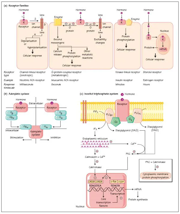
Acromegaly is usually caused by anterior pituitary gland tumours which secrete growth hormone. In 30 to 40% of cases, the tumour is thought to arise due to a somatic mutation affecting transmembrane signalling mechanisms. The stimulatory G-protein Gs is involved in signal transduction at the growth hormone releasing hormone receptor. Mutation of the α-subunit of Gs into the gsp oncogene prolongs the activation phase of the G-protein system, allowing unrestrained hormone synthesis and cell division. The distinctive clinical features of acromegaly and development of the pituitary tumour follow.
Introduction
Hormones interact with target cells through a primary interaction with receptors which recognize the hormones selectively. There are several different receptor systems, which vary in mechanism and timing (Fig. 3a). Charged ions such as peptides and neurotransmitters bind to receptors on the cell membrane. This causes a conformational change in other membrane proteins, which activate enzymes inside the cell, resulting in, for example, the synthesis of ‘second messengers’, which activate phosphorylating enzymes.
Uncharged molecules, such as the steroid hormones diffuse into the cell and bind to intracellular receptors (see Chapter 4). The hormone-receptor complex binds to specific hormone response elements (HRE) on the DNA; the result is that RNA and protein synthesis are altered. The cell will react faster to peptide hormones and neurotransmitters than it will to steroid hormones, which work through relatively slow changes in protein synthesis.Nevertheless, membrane receptors have been discovered for steroid hormones, although the si...