This is the definitive reference for the small animal practitioner to normal radiographic anatomy of the cat and dog. With over forty years of experience between them, the authors have produced an invaluable reference atlas for the veterinary practitioner. The book is suitable for the general and referral based practitioner, undergraduate or postgraduate veterinary surgeon.
Over 550 radiographic images analysed and explained
More than 50 new figures added, with the quality of existing images enhanced
Revised contents and page headers for easy-reference
Clear informative line drawings to trace radiographic shadows and schematic drawings of underlying structures not seen in plain radiographs.
Trusted by 375,005 students
Access to over 1.5 million titles for a fair monthly price.
Mediolateral and the corresponding orthoganol projections.
Additional projections and schematic drawings as indicated.
Hip joints and pelvis, including ‘frog legged’ and oblique, with schematic drawings: Figures 115–128
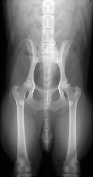
Figure 115Ventrodorsal projection of hip joints and pelvis with full extension of femurs (stifle joints included for hip dysplasia evaluation). Beagle dog 2.5 years old, entire male. (Approximately 60% of original size.)
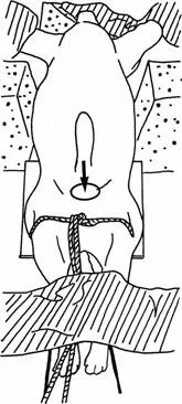
Figure 116 Line drawing of photograph representing radiographic positioning for Figure 115.
Figure 117Ventrodorsal projection of hip joints and pelvis with full extension of femurs. To simplify the labelling each structure has been numbered on one side or the other but not on both sides. Also the vertebral column has not been fully labelled.
A Ilium
1 Crest
2 Gluteal surface
3 Tuber sacrale or dorsal iliac spine
3(a) Cranial dorsal iliac spine
3(b) Caudal dorsal iliac spine
4 Wing
5 Tuber coxae or ventral iliac spine
5(a) Cranial ventral iliac spine
5(b) Caudal ventral iliac spine
6 Body
B Pubis
7 Position of iliopubic eminence. Eminence is often seen as a distinct process where cranial pubic border joins ilium.
8 Pecten
9 Pubic symphysis. Part of symphysis of pelvis.
C Ischium
10 Ischiatic symphysis. Part of symphysis of pelvis.
11 Obturator foramen
12 Ischiatic spine
13 Ischiatic table
14 Ischiatic tuberosity
15 Ischiatic arch
D Acetabulum
16 Cranial acetabular edge
17 Cranial effective acetabular rim
18 Dorsal acetabular edge
19 Ventral acetabular edge
20 Acetabular fossa
20(a) Acetabular notch
20(b) Acetabular fissure
E Femur
21 Head
22 Neck
23 Greater trochanter
23(a) Trochanteric fossa
24 Lesser trochanter (more distinct in left leg on this X-ray)
25 Body
26 Lateral condyle
27 Medial condyle
28 Intercondyloid fossa F Sacrum
29 Wing
30 Lateral sacral crest
31 Median sacral crest
32 Articular surface with ilium wing
32(a) Synovial part of articular surface
32(b) Cartilaginous part of articular surface G Tibia
H Patella
I Fabella of m. gastrocnemius (lateral and medial heads)
J Fabella of m. popliteus
K Coccygeal vertebra
L Lumbar vertebra. (Chronic degenerative changes are present on the left side of 6th and 7th vertebrae at disc space level. Please see ‘Normality’ in the Introduction.)
M Os penis
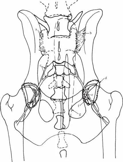
Figure 118 Schematic drawing of ventrodorsal projection of hip joints and pelvis with full extension of femurs to demonstrate extent of joints and ligaments.
Sacroiliac joint
This is a combination of a synovial and cartilaginous joint. The joint capsule is very thin and the two wings are united by a layer of fibrocartilage. Both ventrally and dorsally wide bands of sacroiliac ligaments cover the joint capsule. The dorsal group are more substantial.
a Dorsal sacroiliac ligament
b Ventral sacroiliac ligament
c Sacrotuberous ligament
Hip joint
d Joint capsule
e Ligament of the head of the femur. Formerly called the round ligament. It extends from the fovea capitis of the femoral head to the acetabular fossa. The fovea capitus is not clearly seen in this radiograph but is often visible as a flattening on the medial aspect of the femoral head.
f Transverse acetabular ligament
g Soft tissue shadow of prepuce. This shadow often causes confusion if it is not identified and traced along its entire length. The increase in radiopacity create...
Table of contents
Cover
Title Page
Copyright
Dedication
Preface
Acknowledgements
Introduction
Dog Forelimb
Dog Hindlimb
Dog Skull
Dog Vertebrae, Ribs and Plain radiography Sternum
Dog Pharynx and Larynx
Dog Thorax
Dog Abdomen
Cat Forelimb
Cat Hindlimb
Cat Skull
Cat Vertebrae, Ribs and Sternum
Cat Pharynx, Larynx and Thorax
Cat Abdomen
Dog Soft Tissue
Dog Skeletal System
Cat Soft Tissue
Cat Skeletal System
Bibliography
Frequently asked questions
Yes, you can cancel anytime from the Subscription tab in your account settings on the Perlego website. Your subscription will stay active until the end of your current billing period. Learn how to cancel your subscription
No, books cannot be downloaded as external files, such as PDFs, for use outside of Perlego. However, you can download books within the Perlego app for offline reading on mobile or tablet. Learn how to download books offline
Perlego offers two plans: Essential and Complete
Essential is ideal for learners and professionals who enjoy exploring a wide range of subjects. Access the Essential Library with 800,000+ trusted titles and best-sellers across business, personal growth, and the humanities. Includes unlimited reading time and Standard Read Aloud voice.
Complete: Perfect for advanced learners and researchers needing full, unrestricted access. Unlock 1.5M+ books across hundreds of subjects, including academic and specialized titles. The Complete Plan also includes advanced features like Premium Read Aloud and Research Assistant.
Both plans are available with monthly, semester, or annual billing cycles.
We are an online textbook subscription service, where you can get access to an entire online library for less than the price of a single book per month. With over 1.5 million books across 990+ topics, we’ve got you covered! Learn about our mission
Look out for the read-aloud symbol on your next book to see if you can listen to it. The read-aloud tool reads text aloud for you, highlighting the text as it is being read. You can pause it, speed it up and slow it down. Learn more about Read Aloud
Yes! You can use the Perlego app on both iOS and Android devices to read anytime, anywhere — even offline. Perfect for commutes or when you’re on the go. Please note we cannot support devices running on iOS 13 and Android 7 or earlier. Learn more about using the app
Yes, you can access An Atlas of Interpretative Radiographic Anatomy of the Dog and Cat by Arlene Coulson,Noreen Lewis in PDF and/or ePUB format, as well as other popular books in Medicine & Veterinary Medicine. We have over 1.5 million books available in our catalogue for you to explore.