![]()
1
Cell biology
The cell is the basic unit of living organisms; while some organisms are made up of a single cell (e.g. protozoa and bacteria), others are made up of many cells, organised into tissues and organs, that perform specific functions.
Individual cells in humans and other eukaryotic organisms are organised into functional areas–organelles–that perform a specific function. Cells usually divide by mitosis to produce identical daughter cells to allow the development of tissue or the replacement of dying cells. However, for the purpose of reproduction, they divide by meiosis in which the daughter cells each possess half a full set of chromosomes. In developed tissues, mitosis occurs to replace those cells that have become damaged; if such cell division occurs in an unregulated fashion, cancer may result.
Organelles: structure and function
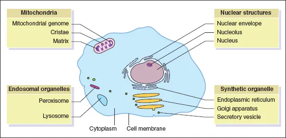
A cell contains specialised regions that take on specific functions. These organelles are discussed below; however, the biochemical interactions that occur are discussed in more detail in Chapter 3.
The cell membrane
The cell membrane is a phospholipid bilayer that surrounds the cell, defining its boundaries. The membrane contains many specialised molecules embedded within it, for the transport of molecules across it, as well as regulating the properties and behaviour of the membrane itself (Fig. 1.1).
Structure
The formation of the bilayer relies on the properties of the constituent phospholipids. They are made up of two parts:
1 A polar head region, which is soluble in water (hydrophilic). This region often contains a charged group which mediates its hydrophilic nature.
2 A non-polar tail region, which is insoluble in water (hydrophobic). This hydrophobic property results from the long uncharged fatty acyl chains.
These properties promote phospholipids to form a bilayer, in which the hydrophilic head regions are in contact with water while the hydrophobic tail regions accumulate in the middle of the bilayer. The fluidity of the membrane is regulated by the presence of cholesterol.
The cell membrane is a dynamic structure, with the various protein components free to rotate and diffuse laterally around the lipids, allowing their aggregation, which is often required for signalling. These proteins can be grouped into two types:
1 Integral proteins are embedded in the membrane.
2 Peripheral proteins are associated with the surface of the membrane as a result of non-covalent interactions.
The precise proteins associated with the membrane differ depending on the cell specialisation, other external signals that have been received and the specific state of the individual cell.
Both proteins and lipids in the membrane may be glycosylated, whereby carbohydrates are added to the molecule. These carbohydrates may be involved in the interaction between the cell and the environment or other cells.
The cytoplasm
The organelles are contained in the cytoplasm, which is made up of a wide variety of ions and solutes, as well as a complex cytoskeletal structure. This structure maintains the cell shape and regulates various transport and trafficking pathways in the cell. The cytoplasm is also the site of most cellular metabolic reactions.
The nucleus
The nucleus contains almost all the genetic information necessary to produce any cell in the human body; this genetic information is densely packed as chromatin. The nucleus is separated from the rest of the cell by a nuclear envelope, which is made up of two lipid bilayers – the outer membrane being continuous with the endoplasmic reticulum. Transport of molecules across the nuclear membrane is tightly regulated by many nuclear pores in the nuclear envelope.
Despite its important function, the nucleus usually occupies a relatively small volume, often around 5% of the total cell volume.
The nucleolus, which is the site of synthesis of ribosomal RNA and the assembly of ribosomes, is one of the few structures visible in the nucleus (by light microscopy).
The endoplasmic reticulum
The endoplasmic reticulum (ER) is a network of tubes continuous with the outer membrane of the nuclear envelopes. It produces lipids and protein for secretion or use in cellular organelles. There are two types of ER:
1 Rough ER is the site of protein production. Its ‘rough’ appearance under an electron microscope comes from the presence of protein-synthesising ribosomes on its surface. The resulting proteins are secreted into the ER lumen, from where they are transported for further modification
2 The smooth ER lacks ribosomes and is responsible for the production of lipids, which contribute to the cellular membranes, and also the production of steroids. Smooth ER is associated with detoxifying reactions, particularly in the liver, and is also the site from which components are transported to other organelles by vesicular transport.
The proportions of rough and smooth ER may reflect the specialisation of the cell, e.g. in the adrenal cortex, which is responsible for the production of steroid hormones, the smooth ER accounts for most of the cell volume.
The Golgi apparatus
The Golgi apparatus modifies many cellular proteins, through the addition of carbohydrates.
It resembles a series of sac-like structures – cisternae – which receive proteins from the rough ER by vesicular transport. As proteins are transported between the different cisternae, they receive carbohydrate modifications; transport may be forwards or backwards between compartments, before the proteins are packaged into vesicles for secretion or transport to specific organelles. This process, trafficking, relies on the presence of specific signals in the proteins themselves, which target them to their final destination.
Mitochondria
Mitochondria synthesise ATP and other phosphate compounds that power cellular reactions through the catabolism of a variety of metabolic compounds.
Structure
Mitochondria are double-membrane organelles. The outer membrane is smooth whereas the inner membrane possesses a series of projections – cristae – that impinge on the interior of the mitochondria, known as the matrix.
Mitochondria contain a primitive genome that encodes some of the proteins not found in the nuclear genome that are necessary for their function. This, and their replication independently of the nucleus of the cell, may reflect their endosymbiotic origin. However, many of the proteins that mitochondria require are solely encoded in the main genome in the nucleus of the cell.
Unlike the main human genome, the mitochondrial genome is always inherited entirely from the mother; mitochondria in sperm do not contribute to the zygote.
DEFINITION Endosymbiotic theory of the origin of mitochondria The mitochondrial genome has been sequenced and has many features consistent with a bacterial genome. It has been suggested that, at some point in the past, a bacterium became engulfed by a primitive cell and remained within it, resulting in a symbiosis between the two organisms. It is thought that this symbiotic bacterium evolved into the mitochondria of today.
Function
Mitochondria meet the energy needs of the cell. In the matrix the TCA (tricarboxylic acid) cycle occurs, which supplies the electron transport chain with the reduced co-factors necessary to generate ATP, the main energy currency of the cell.
The number of mitochondria present in the cell reflects its energy need; muscle cells and neurons have high energy demands and frequently have many mitochondria, whereas cells with lower energy requirements may possess very few.
CLINICAL Myoclonic epilepsy with ragged red fibres
Myoclonic epilepsy with ragged red fibres (MERRF) is a rare condition characterised by myoclonus, epilepsy and ataxia, usually manifesting in the tee...