The aim of the present volume is to provide a more sophisticated text on human oral mucosa than presently exists in textbooks and to bring together information that is otherwise to be found in separate, specialist volumes into a comprehensive text. It relates structure at the molecular, cellular and tissue level to function and to clinical behavior. The volume is directed to advanced students and researchers in oral biology, as well as those in allied areas of investigation, such as dermatology, gynecology, internal medicine and pathology.
- English
- ePUB (mobile friendly)
- Available on iOS & Android
eBook - ePub
About this book
Human Oral Mucosa: Development, Structure and Function is a new text that reflects the considerable increase in knowledge of oral mucosa that has occurred in recent years. Our understanding of the structure of oral mucosa is now established at a molecular rather than a tissue or cellular level. This in turn has revealed a level of function that was previously not suspected, including a sophisticated barrier to the penetration of exogenous materials, and the synthesis of specific antimicrobial compounds, representing components of the innate immune system. There is also a growing realization of commonality in structure and function between regions of oral mucosa and the mucosae of the esophagus and vagina.
Trusted by 375,005 students
Access to over 1.5 million titles for a fair monthly price.
Study more efficiently using our study tools.
Information
1
The functions of oral mucosa
1.1 ORAL MUCOSA: WHAT IS IT AND WHAT DOES IT DO?
Oral mucosa is a mucous membrane, a term used to describe the moist linings of body cavities that communicate with the exterior. These include the oral cavity, nasal passages, pharynx, gastrointestinal tract, and urinogenital regions. In the oral cavity, this lining is called the oral mucous membrane or oral mucosa. The exterior of the body has a dry covering, the skin, which is continuous with oral mucosa at the lips. Structurally, the oral mucosa resembles the skin in some respects and is very similar to the mucous membranes of the esophagus, cervix, and vagina (which will be considered in a subsequent chapter) but is totally different from the gastrointestinal mucosa.
Despite these differences, skin and the different mucosae all consist of two structurally different tissue components: a covering epithelium and an underlying connective tissue. These tissues function together so the various mucosae and skin can be considered as organs.
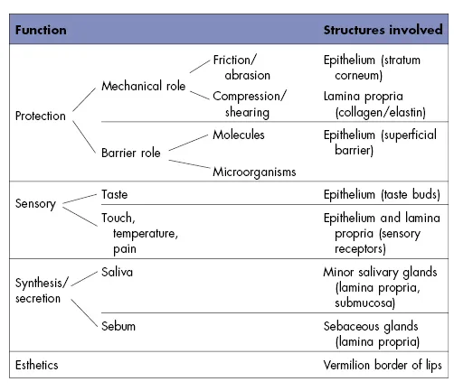
Form follows function, and it is easier to understand the complex structure of a tissue or organ when its function is known. This is particularly true of the oral mucosa, whose structure reflects a variety of functional adaptations. The major adaptations are a result of evolutionary changes that have taken place over a long time. However, small and usually reversible changes in structure of oral mucosa may be seen in response to function during the lifetime of an individual, but these are not heritable. The functions of oral mucosa and the tissue components subserving those functions are summarized in Table 1.1.
Table 1.1 Functions of the oral mucosa.
1.2 FUNCTIONS OF THE ORAL MUCOSA
The oral mucosa has a variety of functions of which the most important is protection of the deeper tissues and glands of the oral cavity. Other functions include sensory perception, synthesis, and secretion from glands located in the mucosa and an esthetic role represented by the mucocutaneous junction.
1.2.1 Protection
As a surface lining, the oral mucosa separates and protects deeper tissues and organs in the oral region from the environment of the oral cavity. The normal activities of seizing, biting, and chewing food expose the oral soft tissues to mechanical forces (compression, stretching, shearing) and surface abrasions (from hard particles in the diet). The oral mucosa shows a number of adaptations of both the epithelium and the connective tissue to withstand these mechanical insults. Furthermore, there is normally a resident population of microorganisms within the oral cavity that would cause infection if they gained access to the tissues. Many of these organisms also produce substances that have a toxic effect on tissues. The epithelium of the oral mucosa acts as the major barrier to penetration and also contributes to the immunoprotective system of the mucosa.
1.2.2 Sensation
The sensory function of the oral mucosa is important because it provides considerable information about events within the oral cavity, whereas the lips and tongue perceive stimuli outside the mouth. In the mouth, pharynx and epiglottis are receptors that respond to temperature, touch, and pain; there also are the taste buds, which are not found anywhere else in the body. These signal the traditional taste sensations of sweet, salty, sour, bitter, and umami (or savory), although it has been suggested recently that there is a “fat” taste (Laugerette et al., 2007). Certain receptors in the oral mucosa probably respond to the “taste” of water and signal the satisfaction of thirst (de Araujo et al., 2003). Reflexes such as swallowing, gagging, retching, and salivating are also initiated by receptors in the oral mucosa.
1.2.3 Secretion
The major secretion associated with the oral mucosa is saliva, produced by the salivary glands, which contributes to the maintenance of a moist surface. The major salivary glands are situated distant from the mucosa, and their secretions pass through the mucosa via long ducts; however, many minor salivary glands are associated with the oral mucosa. Sebaceous glands are frequently present in the oral mucosa, and their secretions may have antimicrobial properties (see Chapter 2). Salivary glands secrete histatins, a family of low-molecular-weight histidine-rich proteins with antimicrobial activities. Oral epithelium is also capable of secreting a variety of antimicrobial factors such as defensins and cathelicidins, which participate in various aspects of innate immunity. These are described in Chapter 8.
1.2.4 Thermal Regulation
In some animals (such as the dog), considerable body heat is dissipated through the oral mucosa by panting; for these animals, the mucosa plays a major role in the regulation of body temperature. The human oral mucosa, however, plays practically no role in regulating body temperature, and there are no obvious specializations of the blood vessels for controlling heat transfer such as arteriovenous shunts.
1.2.5 Esthetics
Skin color, texture, and appearance play an important role in signaling individual characteristics such as age, health, ethnicity, and so on. The oral mucosa is not normally visible except for the region where it joins the skin. Here, the vermilion zone of the lips represents a significant esthetic component, frequently enhanced with cosmetics in females.
REFERENCES
de Araujo, I.E., Kringelbach, M.L., Rolls, E.T., and McGlone, F. (2003) Human cortical responses to water in the mouth, and the effects of thirst. J Neurophysiol 90(3):1865–1876.
Laugerette, F., Gaillard, D., Passilly-Degrace, P., Niot, I., and Besnard, P. (2007) Do we taste fat? Biochimie 89:265–269.
2
The organization of oral mucosa
The oral cavity consists of two parts: an outer vestibule, bounded by the lips and cheeks, and the oral cavity proper, separated from the vestibule by the alveolus bearing the teeth and gingiva. The superior zone of the oral cavity proper is formed by the hard and soft palates, and the floor of the mouth and base of the tongue form the inferior border. Posteriorly, the oral cavity is bounded by the pillars of the fauces and the tonsils. The oral mucosa shows considerable structural variation in different regions of the oral cavity, but three main types of mucosa can be recognized, identified according to their primary function: masticatory mucosa, lining mucosa, and specialized mucosa. The anatomic location of each type is shown diagrammatically in Figure 2.1, and the types are fully described later in the chapter. Quantitatively, the larger part of the oral mucosa is represented by lining mucosa, amounting to approximately 60% of the total area, with masticatory mucosa and specialized mucosa occupying relatively smaller areas.
Figure 2.1 The anatomic locations of the three main types of mucosa in the oral cavity. Masticatory mucosa is shown by black shading; lining mucosa by gray shading; specialized mucosa by the stippled area.
(Modified from Roed-Petersen and Renstrup, 1969, Acta Odontol Scand 27:681.)

2.1 CLINICAL FEATURES
Although the oral mucosa is continuous with the skin, it differs considerably in appearance. Generally it is more deeply colored, most obviously at the lips (where the bright vermilion zone contrasts with the skin tone). This coloration represents the combined effect of a number of factors: the concentration and state of dilation of capillaries in the underlying connective tissue, the thickness of the epithelium, the degree of keratinization, and the amount of melanin pigment in the epithelium. Color gives an indication as to the clinical condition of the mucosa; inflamed tissues are red, because of dilation of the blood vessels, whereas normal healthy tissues are a paler pink (Fig. 2.2A).
Figure 2.2 The oral mucosa lining part of the vestibule. (A) The attached gingiva (G) is pale, and stippling is most evident in the interproximal regions (arrows). There is an abrupt junction (indicated by the dashed line) between the gingival and the alveolar mucosa (AM) which merges with the labial mucosa (LM). (B) Vermilion zone adjoining the labial mucosa. Several small globules on the mucosa (arrows) represent sites of secretion, where minor salivary gland ducts open to the surface.
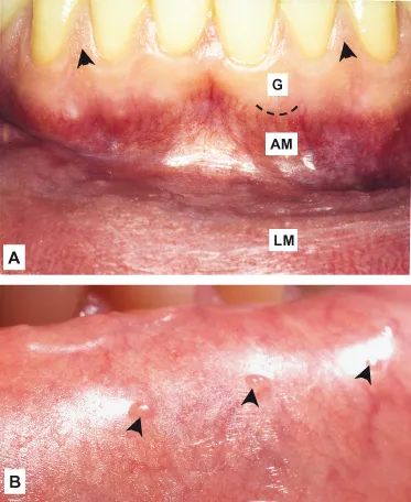
Other features that distinguish the oral mucosa from skin are its moist surface and the absence of appendages. Skin contains numerous hair follicles, sebaceous glands, and sweat glands, whereas the glandular component of oral mucosa is represented primarily by the minor salivary glands. These are concentrated in various regions of the oral cavity, and the openings of their ducts at the mucosal surface are sometimes evident on clinical examination after drying the surface (Fig. 2.2B).
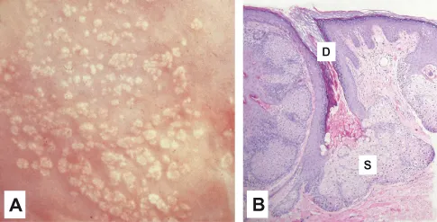
Sebaceous glands are present on the lips, labial mucosa, and buccal mucosa in over three quarters of adults and have been described occasionally in the alveolar mucosa and dorsum of the tongue. They are not associated with hair follicles and are sometimes called sebaceous follicles. Clinically, they appear as pale yellow spots (Fig. 2.3A), sometimes called Fordyce’s spots (or granules) or Fordyce’s disease, although they do not represent a pathologic condition.
Figure 2.3 Sebaceous glands in human buccal mucosa. (A) The “granular” appearance is clearly evident on the buccal mucosal surface. (B) Histological section of a sebaceous gland showing the secretory portion (S) and the duct (D) opening at the surface. (Photograph courtesy of Dr. M.W. Finkelstein.)
The surface of the oral mucosa tends to be smoother and have fewer folds or wrinkles than the skin, but topographic features are readily apparent on clinical examination. The most obvious are the different papillae on the dorsum of the tongue and the transverse ridges (or rugae) of the hard palate. The healthy gingiva shows a pattern of fine surface stippling, consisting of small indentations of the mucosal surface (Fig. 2.2A). In approximately 10% of the population, a slight whitish ridge occurs along the buccal mucosa in the occlusal plane of the teeth. This line, sometimes called the linea alba (white line), is a keratinized region and may represent the epithelial reaction to abrasion from rough tooth restorations or cheek biting.
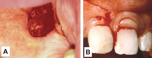
The oral mucosa varies considerably in its firmness and texture. The lining mucosa of the lips and cheeks, for example, is soft and pliable, whereas the gingiva and hard palate are covered by a firm, immobile layer. These differences have important clinical implications when it comes to giving local injections of anesthetics or taking biopsies of oral mucosa. Fluid can be easily introduced into loose lining mucosa, but injection into the masticatory mucosa is more difficult and can be painful for the patient. Lining mucosa gapes when surgically incised (Fig. 2.4A) and frequently requires suturing, whereas masticatory mucosa, being more firmly attached, may not (Fig. 2.4B). Similarly, the accumulation of fluid with inflammation is obvious and painful in masticatory mucosa, but in lining mucosa the fluid disperses, and inflammation may not be so evident or as painful.
Figure 2.4 Wounds in soft palate and gingiva. (A) A small (4 mm) biopsy wound in soft palate, a lining mucosa, results in a wound that has gaped. (B) Incisions in masticatory mucosa (attached gingiva) show little gaping of the wound. (Photograph courtesy of Dr. Georgia Johnson.)
2.2 COMPONENT TISSUES AND GLANDS
The two main tissue components of the oral mucosa are a stratified squamous epithelium, called the oral epithelium, and an underlying connective tissue layer, called the lamina propria (Fig. 2.5). In the skin these two tissues are known by slightly different terminology: epidermis and dermis. The interface between epithelium and connective tiss...
Table of contents
- Cover
- Halftitle page
- Title page
- Copyright page
- Preface
- 1 The functions of oral mucosa
- 2 The organization of oral mucosa
- 3 Oral epithelium
- 4 The interface between epithelium and connective tissue
- 5 Connective tissue
- 6 Regional differences in the oral mucosa
- 7 Development and aging of the oral mucosa
- 8 Barrier functions of oral mucosa
- 9 Homologies in structure and function among mucosae: oral, esophageal, and vaginal mucosa
- Index
Frequently asked questions
Yes, you can cancel anytime from the Subscription tab in your account settings on the Perlego website. Your subscription will stay active until the end of your current billing period. Learn how to cancel your subscription
No, books cannot be downloaded as external files, such as PDFs, for use outside of Perlego. However, you can download books within the Perlego app for offline reading on mobile or tablet. Learn how to download books offline
Perlego offers two plans: Essential and Complete
- Essential is ideal for learners and professionals who enjoy exploring a wide range of subjects. Access the Essential Library with 800,000+ trusted titles and best-sellers across business, personal growth, and the humanities. Includes unlimited reading time and Standard Read Aloud voice.
- Complete: Perfect for advanced learners and researchers needing full, unrestricted access. Unlock 1.5M+ books across hundreds of subjects, including academic and specialized titles. The Complete Plan also includes advanced features like Premium Read Aloud and Research Assistant.
We are an online textbook subscription service, where you can get access to an entire online library for less than the price of a single book per month. With over 1.5 million books across 990+ topics, we’ve got you covered! Learn about our mission
Look out for the read-aloud symbol on your next book to see if you can listen to it. The read-aloud tool reads text aloud for you, highlighting the text as it is being read. You can pause it, speed it up and slow it down. Learn more about Read Aloud
Yes! You can use the Perlego app on both iOS and Android devices to read anytime, anywhere — even offline. Perfect for commutes or when you’re on the go.
Please note we cannot support devices running on iOS 13 and Android 7 or earlier. Learn more about using the app
Please note we cannot support devices running on iOS 13 and Android 7 or earlier. Learn more about using the app
Yes, you can access Human Oral Mucosa by Christopher Squier,Kim Brogden in PDF and/or ePUB format, as well as other popular books in Medicine & Oral Health & Surgery. We have over 1.5 million books available in our catalogue for you to explore.