![]()
1
Anatomical terms
Correct use of anatomical terms is essential to accurate description. These terms are also essential in clinical practice to allow effective communication.
Anatomical position
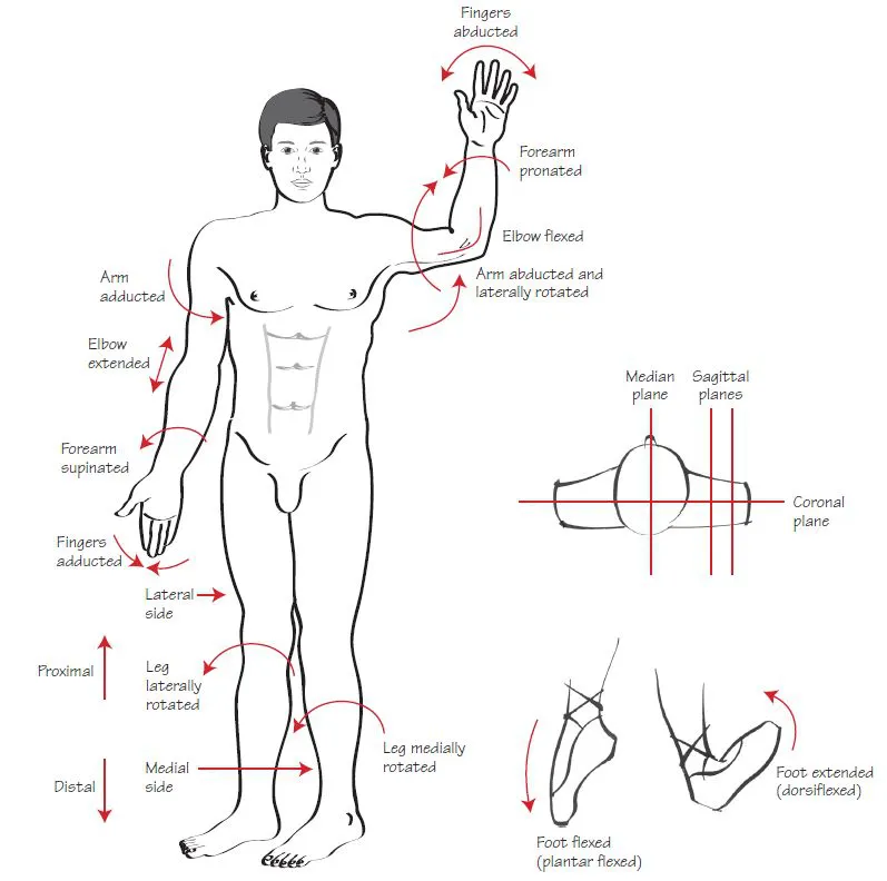
It is important to appreciate that the surfaces of the body, and relative positions of structures, are described, assuming that the body is in the ‘anatomical position’. In this position, the subject is standing upright with the arms by the side with the palms of the hands facing forwards. In the male the tip of the penis is pointing towards the head.
Surfaces and relative positions
- Anterior/posterior: the anterior surface of the body is the front, with the body in the anatomical position. The shin, for example, is referred to as the anterior aspect of the leg, regardless of its position in space. The term ‘posterior’ refers to the back of the body. These terms can also be used to describe relative positions. The bladder, for example, may be described as being anterior to the rectum, or the rectum posterior to the bladder.
- Superior/inferior: these terms refer to vertical relationships in the long axis of the body, between the head and the feet. Superior refers to the head end of the body, inferior to the foot end. These terms are most commonly used to describe relative position. The head, for example, may be described as superior to the neck. It is important to remember that the anatomical position refers to a standing subject. When a patient is lying down, their head remains superior to their neck.
- Medial/lateral: these terms refer to relationships relative to the mid- line of the body. A structure which is medial is nearer the midline, and a lateral structure is further away. So, for example, the inner thigh may be referred to as the medial part of the thigh, and the outer thigh as the lateral part. These terms are also used to describe relationships; the lung may be described as lateral to the heart, or the heart may be described as medial to the lung. In some parts of the body, these terms may cause confusion. The mobility of the forearm in space means that it is easy to get confused about which side is medial or lateral. The terms ‘radial’ and ‘ulnar’, referring to the relationship of the forearm bones, are often used instead.
- Proximal and distal: these terms are used to refer to relationships of structures relative to the middle of the body, the point of origin of a limb or the attachment of a muscle. These terms are commonly used to describe relationships along the length of a limb. A proximal structure is nearer the origin and a distal one further away. The hand is distal to the elbow, for example, and the elbow proximal to the hand.
- Ventral/dorsal: these terms are slightly different from anterior/posterior as they refer to the front and back of the body in terms of embryological development rather than the anatomical position. For the majority of the body, the anterior surface corresponds to the ventral surface and the posterior surface to the dorsal surface. The lower limb is one exception as it rotates during development such that the ventral parts come to lie posteriorly. The ventral surface of the foot, therefore, is the sole.
The ventral surface of the hand is often referred to as the palmar surface and that of the foot as the plantar surface.
- Cranial/caudal: These terms also refer to embryonic development. Cranial refers to the head end of the embryo, and caudal to the tail end.
Planes
Anatomical planes are used to describe sections through the body as if cut all the way through. These planes are essential to understanding cross-sectional imaging:
- Sagittal: this plane lies front to back, such that a sagittal section in the midline would divide the body in half through the nose and the back of the head, continuing downwards.
- Coronal: this plane lies at right angles to the sagittal plane and is parallel to the anterior and posterior surfaces of the body.
- Transverse: this plane lies across the body and is sometimes also referred to as the axial or horizontal plane. A transverse section divides the body across the middle, much like the magician sawing his assistant in half.
Movements
The following anatomical terms are used to describe movement:
- Flexion: is usually taken to mean the bending of a joint, such as bending the elbow or knee. Strictly, it refers to the apposition of two ventral surfaces, which is generally taken to mean the same thing.
- Extension: is the straightening of a joint or the movement of two ventral surfaces such that they come to lie further apart.
- Abduction: is movement of a part of a body away from the midline in the coronal plane. For example, abduction of the arm is lifting the arm out sideways.
In the hand, the midline is considered to be along the middle finger. Thus, abduction of the fingers refers to the motion of spreading them out. In the foot, the axis of abduction is the second toe.
The thumb is a special case. Abduction of the thumb refers to anterior movement away from the palm (see Fig 1.1). Adduction is the opposite of this movement.
- Adduction: is movement towards the middle of the body in the coronal plane.
- Plantar/dorsiflexion: are used to describe movement of the foot at the ankle as the use of the terms ‘flexion’ and ‘extension’ is confusing. True flexion of the foot is straightening at the ankle, because this leads to two ventral surfaces coming closer together. This is, however, somewhat confusing. For this reason, the term ‘plantar/flexion’ is used to refer to the action of pointing the toes and dorsiflexion to refer to bending at the ankle such that the toes move towards the face.
- Rotation: rotation is movement around the long axis of a bone. For example rotation of the femur at the hip joint will cause the foot to point laterally or medially.
- Supination/pronation: are special terms used to refer to rotational movements of the forearm, best thought of when the elbow is flexed to 90 degrees. Supination refers to rotation of the forearm at the elbow laterally, such that the palm faces superiorly. Pronation refers to an inward rotation, such that the dorsal surface of the hand is uppermost.
![]()
2
Embryology
Normal pregnancy lasts 40 weeks. The first 8 weeks are termed the embryonic period, during which the body structures and organs are formed and differentiated. The fetal period runs from eight weeks to birth and involves growth and maturation of these structures.
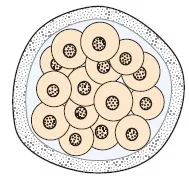
The combination of ovum and sperm at fertilisation produces a zygote. This structure further divides to produce a ball of cells called the morula (Fig. 2.1), which develops into the blastocyst during the 4th and 5th days of pregnancy.
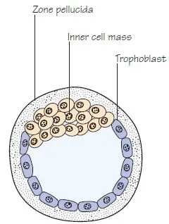
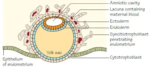
The blastocyst (Fig. 2.2): consists of an outer layer of cells called the trophoblast which encircles a fluid filled cavity. The trophoblast eventually forms the placenta. A ball of cells called the inner cell mass is attached to the inner surface of the trophoblast and will eventually form the embryo itself. At about six days of gestation, the blastocyst begins the process of implanting into the uterine wall. This process is complete by day 10.
Further division of the inner cell mass during the second week of development causes a further cavity to appear, the amniotic cavity. The blastocyst now consists of two cavities, the amniotic cavity and the yolk sac (derived from the original blastocyst cavity) (Fig. 2.3). These cavities are separated by the embryonic plate. The embryonic plate consists of two layers of cells, the ectoderm lying in the floor of the amniotic cavity and the endoderm lying in the roof of the yolk sac.
Gastrulation: is the process during the third week of gestation during which the two layers of embryonic plate divide into three, giving rise to a trilaminar disc. This is achieved by the development of the primitive streak as a thickening of the ectoderm. Cells derived from the primitive streak invaginate and migrate between the ectoderm and endoderm to form the mesoderm. The embryonic plate now consists of three layers:
Ectoderm: eventually gives rise to the epidermis, nervous system, anterior pituitary gland, the inner ear and the enamel of the teeth.
Endoderm: gives rise the epithelial lining of the respiratory and gastrointestinal tracts.
Mesoderm: lies between the ectoderm and endoderm and gives rise to the smooth and striated muscle of the body, connective tissue, blood vessels, bone marrow and blood cells, the skeleton, reproductive organs and the urinary tract.
The notochord and neural plate
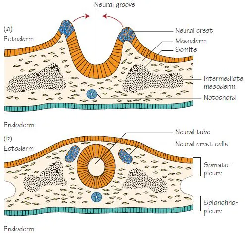
The notochord develops from a group of ectodermal cells in the midline and eventually forms a tubular structure within the mesodermal layer of the embryo. The notochord induces development of the neural plate in the overlying ectoderm and eventually disappears, persisting only in the intervertebral discs as the nucleus pulposus.
The neural plate invaginates centrally to form a groove and then folds to form a tube by the end of week three, a process known as neurulation (Fig. 2.4). The neural tube then becomes incorporated into the embryo, such that it comes to lie deep to the overlying ectoderm. The resultant neural tube develops into the brain and spinal cord.
Some cells from the edge of the neural plate become separated and come to lie above and lateral to the neural tube, when they become known as neural crest cells. These important cells give rise to several structures including the dorsal root ganglia of spine nerves, the ganglia of the autonomic nervous system, Schwann cells, meninges, the chromaffin cells of the adrenal medulla, parafollicular cells of the thyroid and the bones of the skull and face.
Mesoderm
The mesodermal layer of the embryo comes to lie alongside the notochord and neural tube and is subdivided into three parts:
Paraxial mesoderm: lies nearest the midline and becomes segmented into paired clumps of cells called somites. The somites are further divided into the sclerotome, which eventually surrounds the neural tube and notochord to produce the vertebral column and ribs, and the dermatomyotome which forms the muscles of the body wall and the dermis of the skin. The segmental arrangement of the somites explains the eventual arrangement of dermatomes in the body wall and limbs (Fig. 78.1).
Intermediate mesoderm: lies lateral to the paraxial mesoderm. It eventually gives rise to the precursors of the urinary tract(see Chapter 31).
Lateral mesoderm: is involved with the formation of body cavities and the folding of the embryo (Fig. 2.4b).
A separate group of cells from the primitive streak migrate around the neural plate to form the cardiogenic mesoderm, which eventually gives rise to the heart.
Folding of the embryo
The folding of the embryo commences at the beginning of the fourth week (Fig. 2.5). The flat embryonic disc folds as a result of faster growth of the ectoderm cranio-caudally, such that it is concave towards the yolk sac and convex towards the amnion. Lateral folding occurs around the yolk sac in the same manner.
During this process, the lateral plate mesoderm splits to create the embryonic coelom or body cavity (Fig. 2.4). The inner layer is called the splanchnopleure and surrounds the yolk sac in such a way that it becomes incorporated into the embryo, forming the cells lining the lumen of the gastrointestinal tract. The cranial part of the yolk sac migrates further cranially, forming the foregut, and the caudal part migrates further caudally, forming the hindgut (Fig. 2.6). As the folding of the embryo continues the yolk sac forms a small vesicle lying outside the embryo and connected to the gut by a narrow vitello-intestinal duct (see Chapter 31). The two ends of the primitive gut are separated from the amniotic cavity at the cranial end by the buccopharyngeal membrane, and the caudal end by the cloacal membrane, which are formed of ectoderm and endoderm with no intervening mesoderm. They eventually disappear to form cranial and caudal openings into the pharynx and the anal canal, respectively.
The outer layer of the lateral mesoderm is called the somatopleure. This layer is invaded by paraxial mesod...