Imaging in Endocrinology
eBook - ePub

Imaging in Endocrinology

  1. English
  2. ePUB (mobile friendly)
  3. Available on iOS & Android
eBook - ePub

About this book

Imaging in Endocrinology will provide endocrinologists and radiologists of all levels with an outstanding diagnostic imaging atlas to aid them in the diagnosis and management of all the major endocrine diseases they are likely to encounter.

In full colour throughout, the 300 high-quality images consist of CT scans, MRI, NMR and histopathology slides, and are arranged by each specific endocrine condition, resulting in a visually outstanding and easily accessible tool that guides the user through exactly what to look out for and provides a practical and extremely useful aid in helping them formulate a diagnosis. 

Every major endocrine condition is covered in a specific section, including diseases of the thyroid, pituitary, reproductive and adrenal glands, the pancreas, bone metabolism problems, and the various forms of endocrine cancers.  Each disease covered will offer a comparison of the normal findings so as to further assist in diagnosis.  An accompanying website contains an online slide-atlas of all the figures in the book, to allow users to download all figures for use in presentations.

Led by Paolo Pozzilli, an internationally-recognised expert in this field, the authors have assembled a wonderful collection of images that will be greatly valued by endocrinologists and radiologists alike, ensuring this is the perfect tool to consult when assessing patients with endocrine disease.

Frequently asked questions

Yes, you can cancel anytime from the Subscription tab in your account settings on the Perlego website. Your subscription will stay active until the end of your current billing period. Learn how to cancel your subscription.
At the moment all of our mobile-responsive ePub books are available to download via the app. Most of our PDFs are also available to download and we're working on making the final remaining ones downloadable now. Learn more here.
Perlego offers two plans: Essential and Complete
  • Essential is ideal for learners and professionals who enjoy exploring a wide range of subjects. Access the Essential Library with 800,000+ trusted titles and best-sellers across business, personal growth, and the humanities. Includes unlimited reading time and Standard Read Aloud voice.
  • Complete: Perfect for advanced learners and researchers needing full, unrestricted access. Unlock 1.4M+ books across hundreds of subjects, including academic and specialized titles. The Complete Plan also includes advanced features like Premium Read Aloud and Research Assistant.
Both plans are available with monthly, semester, or annual billing cycles.
We are an online textbook subscription service, where you can get access to an entire online library for less than the price of a single book per month. With over 1 million books across 1000+ topics, we’ve got you covered! Learn more here.
Look out for the read-aloud symbol on your next book to see if you can listen to it. The read-aloud tool reads text aloud for you, highlighting the text as it is being read. You can pause it, speed it up and slow it down. Learn more here.
Yes! You can use the Perlego app on both iOS or Android devices to read anytime, anywhere — even offline. Perfect for commutes or when you’re on the go.
Please note we cannot support devices running on iOS 13 and Android 7 or earlier. Learn more about using the app.
Yes, you can access Imaging in Endocrinology by Paolo Pozzilli,Andrea Lenzi,Bart L. Clarke,William F. Young, Jr. in PDF and/or ePUB format, as well as other popular books in Medicine & Endocrinology & Metabolism. We have over one million books available in our catalogue for you to explore.

Information

1
Thyroid
Hashimoto’s thyroiditis (chronic autoimmune thyroiditis)
Definition and epidemiology
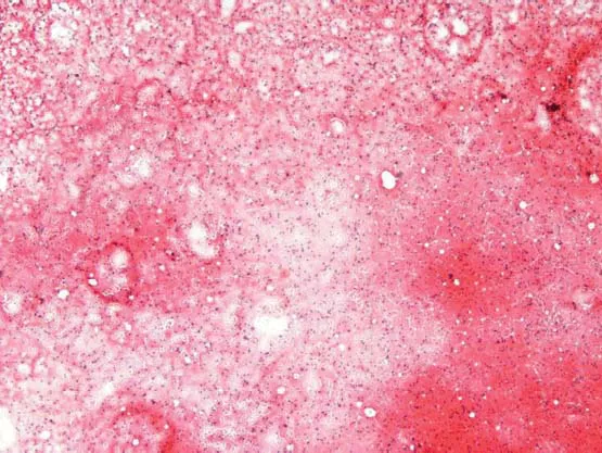
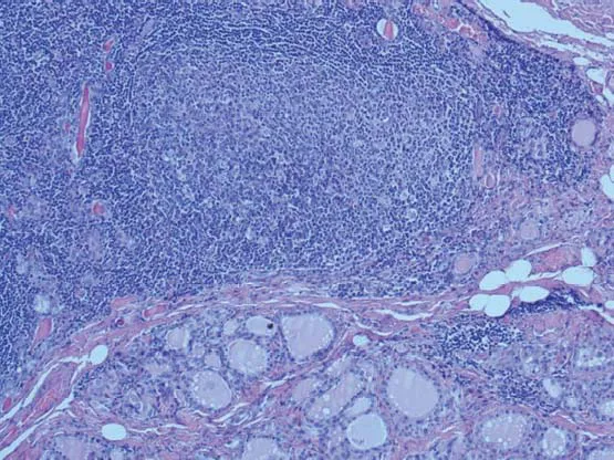
Hashimoto’s thyroiditis (HT), or chronic lymphocytic thyroiditis, is an autoimmune disease in which the thyroid gland is attacked by a variety of cell and antibody-mediated immune processes. The name “Hashimoto’s thyroiditis” is derived from the 1912 original report by Hashimoto describing patients with both goiter and intense lymphocytic infiltration of the thyroid (Figs 1.1 & 1.2) as “struma lymphomatosa.”
Hashimoto’s thyroiditis is the most common cause of primary hypothyroidism in iodine-sufficient areas of the world; it is among the most common causes of nonendemic goiter. On average 1.0–1.5/1000 people suffer from this disease. It occurs far more often in women than in men (incidence of 10 : 1 to 20 : 1, respectively), and it is most prevalent between 45 and 65 years of age. Occurrence in children is also uncommon, especially in populations where iodine is not a dietary scarcity.
Etiology and pathogenesis
Autoantibodies may be present against thyroid peroxidase, thyroglobulin, and thyroid-stimulating hormone (TSH) receptors, although a small percentage of patients may have none of these antibodies present. Antibody-dependent cell-mediated cytotoxicity is a substantial factor behind the apoptotic fallout of HT. Activation of cytotoxic T lymphocytes (CD8+ T cells) in response to cell-mediated immune response affected by helper T lymphocytes (CD4+ T cells) is central to thyrocyte destruction. Recruitment of macrophages is another effect of helper T-lymphocyte activation, with Th1-axis lymphocytes producing inflammatory cytokines within the thyroid tissue to further macrophage activation and migration into the thyroid gland for a direct effect. Infection, stress, sex steroids, pregnancy, iodine intake, and radiation exposure are known possible precipitating factors for HT. Fetal microchimerism within the maternal thyroid is also a possibility.
Signs and symptoms
Hashimoto’s thyroiditis very often results in hypothyroidism with bouts of hyperthyroidism. Symptoms of HT include weight gain, depression, mania, sensitivity to heat and cold, paresthesia, fatigue, panic attacks, bradycardia, tachycardia, high cholesterol, reactive hypoglycemia, constipation, migraine, muscle weakness, cramps, memory loss, infertility, hair loss, and myxedematous psychosis.
Diagnosis
Laboratory findings
Laboratory tests for HT include:
  • Antithyroid peroxidase antibodies (TPOAbs) and thyroglobulin antibodies (TgAbs)
  • TSH, free thyroxine (FT4)
  • Total cholesterol, high density lipoprotein (HDL), and triglycerides
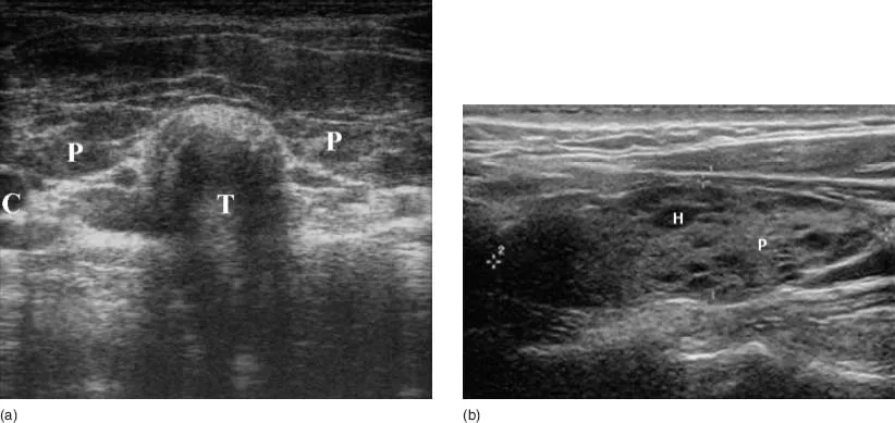
Imaging tests
Imaging tests for HT include:
  • Neck ultrasound (Fig. 1.3)
  • Computed tomography (CT) scan (rare)
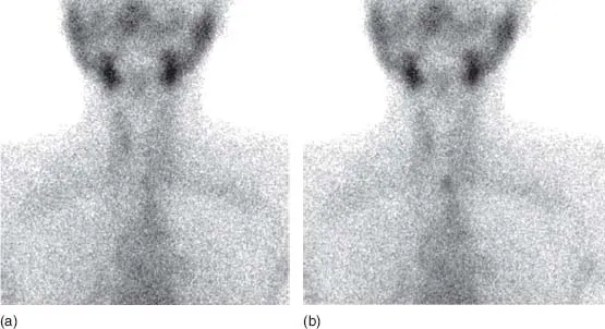
  • 99mTcO4 thyroid scintigraphy (Fig. 1.4)
Treatment
In patients with primary hypothyroidism, the main treatment is levothyroxine.
Illustrations (Figs 1.1–1.4)
Figure 1.1 Cytology of thyroiditis. This figure shows rare and normal thyrocytes associated with numerous lymphocytes (Papanicolau, 10×).
c01f001
Figure 1.2 Histology of thyroiditis. Hashimoto thyroiditis is characterized by Hürthle cells associated with follicular lymphoid structures (HE, 10×).
c01f002
Figure 1.3 A 46-year-old woman with a recent episode of cervical tenderness and a familiar history of thyroid disease. The patient complained of fatigue and reported a weight gain of about 10 kg in the last 2 months. (a) Thyroid ultrasound – cross section. This ultrasound shows a thyroid with a slight increase in volume, globular shape, and homogeneous structure, and less echogenic than normal. (b) Thyroid ultrasound – longitudinal section. This ultrasound shows diffuse patchy hypoechoic lesions throughout the gland. This sonographic appearance is called a “leopard skin” pattern and is seen in lymphocytic infiltration of the thyroid in Hashimoto’s thyroiditis. The hypoechoic lesions within the thyroid are areas of lymphocytic infiltration of the thyroid tissue. C, carotid artery; H, hypoechoic lesions; P, thyroid parenchyma; T, trachea.
c01f003
Figure 1.4 The same patient as in Fig. 1.3: 99mTcO4 thyroid scintigraphy with iodine uptake curve. Iodine uptake was 2% at 4 hours (a) and 2% at 24 hours (b). The scan showed no uptake in the thyroid bed. The free triiodothyronine (FT3) and free thyroxine (FT4) levels were low with elevated thyroid stimulating hormone (TSH) and antibodies against thyroperoxidase (TPOAb) values. The patient started levothyroxine treatment.
c01f004
Graves’ disease (Basedow’s disease)
Definition and epidemiology
Graves’ disease (GD) is an autoimmune disease representing the most common cause of hyperthyroidism (60–90% of all cases).
Graves’ disease has a powerful hereditary component, affecting up to 2% of the female population, and is between five and ten times more common in females than in males (incidence of 5 : 1 to 10 : 1, respectively). It is also the most common cause of severe hyperthyroidism, which is accompanied by extended clinical signs and symptoms and laboratory abnormalities compared with milder forms of hyperthyroidism. About 30–50% of patients with GD will also suffer from Graves’ ophthalmopathy, which is caused by inflammation of the eye muscles mediated by an inflammatory immune process.
Etiology and pathogenesis
Graves’ disease is an autoimmune disorder in which the body produces antibodies to the receptor for thyroid stimulating hormone (TSHrAb). (Antibodies to thyroglobulin and thyroperoxidase may also be produced.) TSHrAb bind to the thyroid stimulating hormone (TSH) receptors, which are located on cells...

Table of contents

  1. Cover Page
  2. Title Page
  3. Copyright Page
  4. About the Companion Website
  5. Preface
  6. Collaborators
  7. Chapter 1: Thyroid
  8. Chapter 2: Pituitary Gland
  9. Chapter 3: Adrenal Gland
  10. Chapter 4: Pancreas
  11. Chapter 5: Bone and Mineral Metabolism
  12. Chapter 6: Gonads
  13. Chapter 7: Mucocutaneous Manifestations of Endocrine Disorders
  14. Index