
- English
- ePUB (mobile friendly)
- Available on iOS & Android
eBook - ePub
Neuroanatomy and Neuroscience at a Glance
About this book
Neuroanatomy and Neuroscience at a Glance provides a user-friendly introduction to the anatomy, biochemistry, physiology and pharmacology of the human nervous system within one, succinct, highly-illustrated volume.
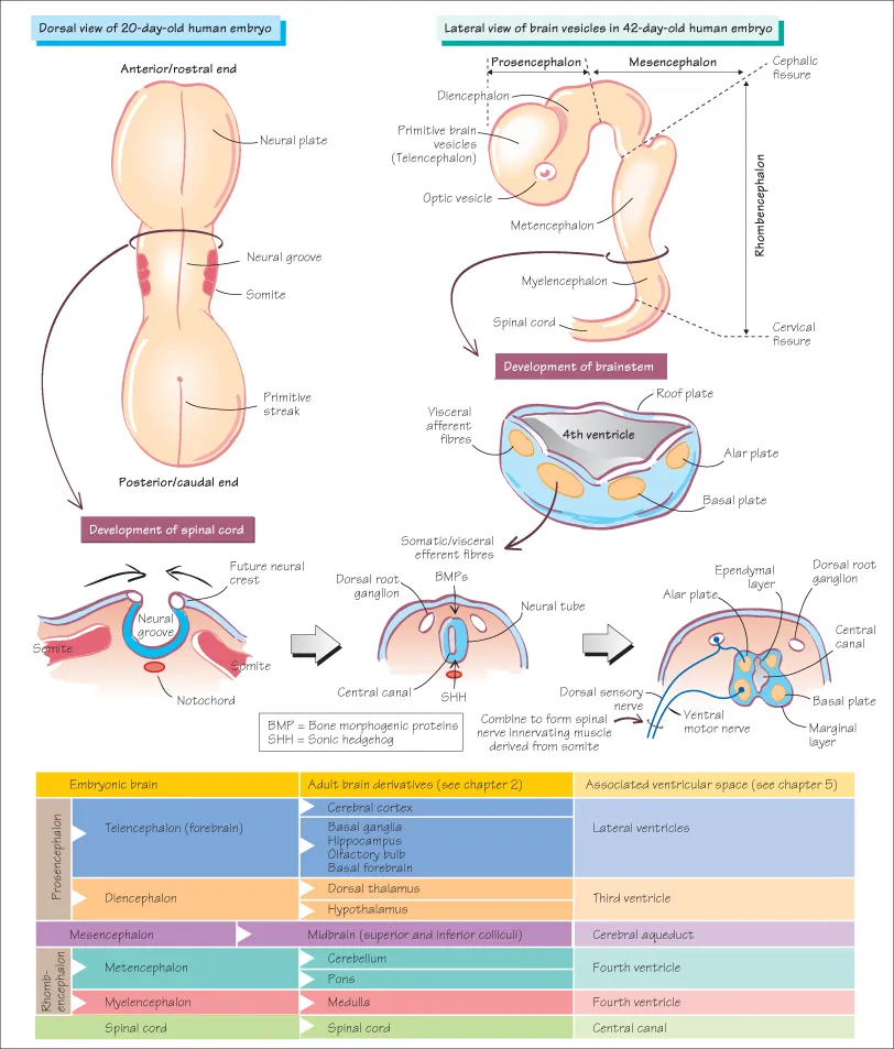
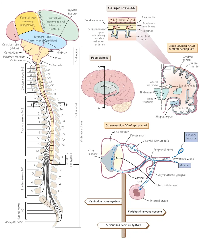
The double page spreads begin by summarising the anatomical structure and function of the different components of the central nervous system, followed by a section on applied neurobiology which outlines how to approach the patient with neurological and psychiatric problems and provides an overview of treatment and management options.
Key features of this fourth edition include:
- A manageable overview of the structure and function of the central nervous system
- Full guidance on how to approach the patient with neurological problems and the investigations used in the most common scenarios
- Cases highlighting the clinical relevance of the basic neuroscience
- New chapters on the major neurotransmitters of the CNS and their functions, the enteric nervous system and stroke
- A fully updated companion website with interactive self-assessment questions and case studies, flashcards and revision notes at www.ataglanceseries.com/neuroscience
Neuroanatomy and Neuroscience at a Glance is the ideal companion for anyone about to start a basic neuroanatomy or neuroscience course, or can be used as a refresher for those in clinical training.
Frequently asked questions
Yes, you can cancel anytime from the Subscription tab in your account settings on the Perlego website. Your subscription will stay active until the end of your current billing period. Learn how to cancel your subscription.
No, books cannot be downloaded as external files, such as PDFs, for use outside of Perlego. However, you can download books within the Perlego app for offline reading on mobile or tablet. Learn more here.
Perlego offers two plans: Essential and Complete
- Essential is ideal for learners and professionals who enjoy exploring a wide range of subjects. Access the Essential Library with 800,000+ trusted titles and best-sellers across business, personal growth, and the humanities. Includes unlimited reading time and Standard Read Aloud voice.
- Complete: Perfect for advanced learners and researchers needing full, unrestricted access. Unlock 1.4M+ books across hundreds of subjects, including academic and specialized titles. The Complete Plan also includes advanced features like Premium Read Aloud and Research Assistant.
We are an online textbook subscription service, where you can get access to an entire online library for less than the price of a single book per month. With over 1 million books across 1000+ topics, we’ve got you covered! Learn more here.
Look out for the read-aloud symbol on your next book to see if you can listen to it. The read-aloud tool reads text aloud for you, highlighting the text as it is being read. You can pause it, speed it up and slow it down. Learn more here.
Yes! You can use the Perlego app on both iOS or Android devices to read anytime, anywhere — even offline. Perfect for commutes or when you’re on the go.
Please note we cannot support devices running on iOS 13 and Android 7 or earlier. Learn more about using the app.
Yes, you can access Neuroanatomy and Neuroscience at a Glance by Roger A. Barker,Francesca Cicchetti in PDF and/or ePUB format, as well as other popular books in Medicine & Neurology. We have over one million books available in our catalogue for you to explore.
Information
Part 1:
Anatomical and functional organization
1
Development of the nervous system

The first signs of nervous system development occur in the third week of gestation, under the influence of secreted factors from the notochord, with the formation of a neural plate along the dorsal aspect of the embryo. This plate broadens, folds (forming the neural groove) and fuses to form the neural tube, which ultimately gives rise to the brain at its rostral (i.e. towards the head) end and the spinal cord caudally (i.e. towards the feet/tail end). The fusion begins approximately halfway along the neural groove at the level of the fourth somite and continues caudally and rostrally with the closure of the posterior/caudal and anterior/rostral neuropore during the fourth week of gestation.
The Development of the Spinal Cord
The process of neural tube fusion isolates a group of cells termed the neural crest.
- The neural crest gives rise to a range of cells including the dorsal root ganglia (DRG) and peripheral components of the autonomic nervous system (ANS; see Chapter 3).
- The DRG contain the sensory cell bodies which send their developing axons into the evolving spinal cord and skin.
- These growing neuronal processes or neurites have an advancing growth cone that finds its appropriate target in the periphery and central nervous system (CNS), using a number of cues including cell adhesion molecules and diffusible neurotrophic factors (see Chapter 48).
The neural tube surrounds the neural canal, which forms the central canal of the fully developed spinal cord.
- The tube itself contains the neuroblasts (ependymal layer), which divide and migrate out to the mantle layer, where they differentiate into neurones to form the grey matter of the spinal cord (see Chapter 2).
- The developing processes from the neuroblasts/neurones grow out into the marginal layer, which therefore ultimately forms the white matter of the spinal cord.
- The dividing neuroblasts segregate into two discrete populations, the alar and basal plates, which in turn will create the dorsal and ventral horns of the spinal cord while a small lateral horn of visceral efferent neurones (part of the ANS) develops at their interface in the thoracic and upper lumbar cord (see Chapter 3).
- This dorso-ventral patterning relies, at least in part, on factors secreted dorsally (bone morphogenic proteins (BMPs)) or ventrally from the notochord (sonic hedgehog (SHH)).
The Development of the Brain
Adult Neurogenesis
Until recently it was believed that no new neurones could be born in the adult mammalian brain. However, it is now clear that neural progenitor cells can be found in the adult CNS, including in humans. These cells are predominantly found in the dentate gyrus of the hippocampus (see Chapter 45) and just next to the lateral ventricles in the subventricular zone (SVZ). They may also exist at other sites of the adult CNS but this is contentious. They respond to a number of signals and appear to give rise to functional neurones in the hippocampus and olfactory bulb, with the latter cells migrating from the SVZ to the olfactory bulb via the rostral migratory stream (RMS). They may therefore fulfil a role in certain forms of memory and possibly in mediating the therapeutic effects of some drugs such as antidepressants (see Chapter 57).
Disorders of Central Nervous System Embryogenesis
- Anencephaly occurs when there is failure of fusion of the anterior rostral neuropore. The cerebral vesicles fail to develop and thus there is no brain formation. The vast majority of fetuses with this abnormality are spontaneously aborted.
- Spina bifida refers to any defect at the lower end of the vertebral column and/or spinal cord. The most common form of spina bifida refers to a failure of fusion of the dorsal parts of the lower vertebrae (spina bifida occulta). This can be associated with defects in the meninges and neural tissue which may herniate through the defect to form a meningocoele and meningomyelocoele, respectively. The most serious form of spina bifida is when nervous tissue is directly exposed as a result of a failure in the proper fusion of the posterior/caudal neuropore. Spina bifida is often associated with hydrocephalus (see Chapter 5). Occasionally, bony defects are found at the base of the skull with the formation of a meningocoele. However, unlike the situation at the lower spinal cord, these can often be repaired without any neurological deficit being accrued.
- Cortical dysplasia refers to a spectrum of defects that are the result of the abnormal migration of developing cortical neurones. These defects are becoming increasingly recognized with improved imaging of the human CNS, and are now known to be an important cause of epilepsy (see Chapter 61).
- Many intrauterine infections (such as rubella), as well as some environmental agents (e.g. radiation), cause major problems in the development of the nervous system. In addition, a large number of rare genetic conditions are associated with defects of CNS development, but these lie beyond the scope of this book.
Did you know?
The adult human brain continues to make new nerve cells throughout life and that this can be promoted by a whole range of activities including exercising, learning new skills and, for example, socializing.
2
Organization of the nervous system

The nervous system can be divided into three major parts: the autonomic (ANS), peripheral (PNS) and central (CNS) nervous systems. The PNS is defined as those nerves that lie outside the brain, brainstem or spinal cord, while the CNS embraces those cells that lie within these structures.
Autonomic Nervous System
- The ANS has both a central and peripheral component and is involved with the innervation of internal and glandular organs (see Chapter 3): it has an important role in the control of the endocrine and homoeostatic systems of the body (see Chapter 3, 11). The peripheral component of the ANS is defined in terms of the enteric (see Chapter 4), sympathetic and parasympathetic systems ...
Table of contents
- Cover
- Table of Contents
- Introduction
- Part 1: Anatomical and functional organization
- Part 2: Cells and neurophysiology
- Part 3: Sensory systems
- Part 4: Motor systems
- Part 5: Consciousness and higher brain function
- Part 6: Applied neurobiology: the principles of neurology and psychiatry
- Part 7: Self-assessment case studies
- Index
- End User License Agreement