Elements of Modern X-ray Physics
eBook - ePub

Elements of Modern X-ray Physics

Jens Als-Nielsen, Des McMorrow

Share book
  1. English
  2. ePUB (mobile friendly)
  3. Available on iOS & Android
eBook - ePub

Elements of Modern X-ray Physics

Jens Als-Nielsen, Des McMorrow

Book details
Book preview
Table of contents
Citations

About This Book

Eagerly awaited, this second edition of a best-selling text comprehensively describes from a modern perspective the basics of x-ray physics as well as the completely new opportunities offered by synchrotron radiation. Written by internationally acclaimed authors, the style of the book is to develop the basic physical principles without obscuring them with excessive mathematics.

The second edition differs substantially from the first edition, with over 30% new material, including:

  • A new chapter on non-crystalline diffraction - designed to appeal to the large community who study the structure of liquids, glasses, and most importantly polymers and bio-molecules
  • A new chapter on x-ray imaging - developed in close cooperation with many of the leading experts in the field
  • Two new chapters covering non-crystalline diffraction and imaging
  • Many important changes to various sections in the book have been made with a view to improving the exposition
  • Four-colour representation throughout the text to clarify key concepts
  • Extensive problems after each chapter

There is also supplementary book materialfor this title available online (http://booksupport.wiley.com).

Praise for the previous edition:

"The publication of Jens Als-Nielsen and Des McMorrow's Elements of Modern X-ray Physics is a defining moment in the field of synchrotron radiation... a welcome addition to the bookshelves of synchrotron–radiation professionals and students alike.... The text is now my personal choice for teaching x-ray physics...."
— Physics Today, 2002

Frequently asked questions

How do I cancel my subscription?
Simply head over to the account section in settings and click on “Cancel Subscription” - it’s as simple as that. After you cancel, your membership will stay active for the remainder of the time you’ve paid for. Learn more here.
Can/how do I download books?
At the moment all of our mobile-responsive ePub books are available to download via the app. Most of our PDFs are also available to download and we're working on making the final remaining ones downloadable now. Learn more here.
What is the difference between the pricing plans?
Both plans give you full access to the library and all of Perlego’s features. The only differences are the price and subscription period: With the annual plan you’ll save around 30% compared to 12 months on the monthly plan.
What is Perlego?
We are an online textbook subscription service, where you can get access to an entire online library for less than the price of a single book per month. With over 1 million books across 1000+ topics, we’ve got you covered! Learn more here.
Do you support text-to-speech?
Look out for the read-aloud symbol on your next book to see if you can listen to it. The read-aloud tool reads text aloud for you, highlighting the text as it is being read. You can pause it, speed it up and slow it down. Learn more here.
Is Elements of Modern X-ray Physics an online PDF/ePUB?
Yes, you can access Elements of Modern X-ray Physics by Jens Als-Nielsen, Des McMorrow in PDF and/or ePUB format, as well as other popular books in Naturwissenschaften & Kondensierte Materie. We have over one million books available in our catalogue for you to explore.

Information

Publisher
Wiley
Year
2011
ISBN
9781119970156
Chapter 1
X-rays and their interaction with matter
X-rays were discovered by Wilhelm Conrad Röntgen in 1895. Since that time they have become established as an invaluable probe of the structure of matter. The range of materials for which X-rays have proved to be decisive in unravelling the structure is truly staggering. These include at one limit of complexity simple compounds, through to more complex and celebrated examples, such as DNA. In more recent times the structure of proteins, and even functional units of living organisms, can be solved on a regular basis. Progress in both our theoretical understanding of the interaction of X-rays with matter, and in our knowledge of how to exploit them experimentally, was steady from the period covering their discovery through to the mid 1970s. The main limitation in this period was the source, which had remained essentially unchanged from about 1912. In the 1970s it was realized that the synchrotron radiation emitted from charged particles circulating in storage rings constructed for high energy nuclear physics experiments was potentially a much more intense and versatile source of X-rays. Indeed synchrotrons have proven to be such vastly better sources that many storage rings have been constructed around the world dedicated solely to the production of X-rays.
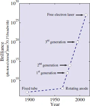
This has culminated to date in the so-called third-generation synchrotron sources, which are more brilliant than the early lab-based sources by a factor of approximately 1012, as indicated in Fig. 1.1. With the advent of synchrotron sources the pace of innovation in X-ray science increased markedly (though perhaps not a trillion fold!), and today shows no signs of slowing. The first X-ray free-electron lasers have recently come into service, and when they become fully operational further important breakthroughs will undoubtedly follow. In Chapter 2 we explain the basic physical principles of X-ray sources and outline their salient properties.
Fig. 1.1 The brilliance of X-ray sources as a function of time. Source brilliance is defined and discussed in Chapter 2, along with the principles underlying the production of X-rays from synchrotrons and free-electron lasers. For free-electron laser sources we plot the average brilliance. Due to the extremely short X-ray pulse length from a free-electron laser − of order 100 fs − the peak brilliance exceeds the average brilliance by a large factor.
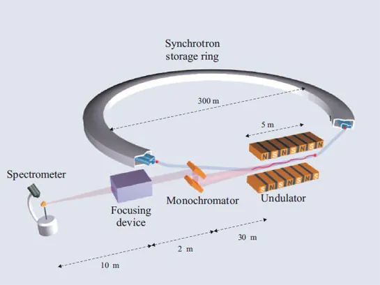
In Fig. 1.2 we show a schematic of the key components of a typical experimental beamline at a third-generation source. The details will of course vary considerably depending on the particular requirements, but many of the components shown will be found in one form or another on most beamlines. First there is the source itself. In this case the electrons do not follow a purely circular orbit in the storage ring, but traverse through straight sections where lattices of magnets, so-called undulator insertion devices, force them to execute small-amplitude oscillations. At each oscillation X-rays are emitted and, if the amplitude of the oscillations is small, then the different contributions from the passage of a single electron add coherently, and a very intense beam of X-rays results. The second key component is the monochromator, as in many applications it is required to work at a particular average wavelength. It may also be desirable to choose the wavelength bandwidth, and monochromators made from perfect crystals through to multilayers allow for a considerable variation in this parameter. Thirdly, if working with small samples it may be desirable to focus the monochromatic beam down to as small a size as achievable. This is accomplished by devices such as X-ray mirrors and refractive Fresnel lenses. Finally, X-rays are delivered to the sample itself on which the experiment is performed.
Fig. 1.2 A schematic of a typical X-ray beamline at a third generation X-ray source. Bunches of charged particles (electrons or positrons) circulate in a storage ring (typical diameter around 300 m). The ring is designed with straight sections, where an insertion device, such as undulator, is placed. The lattice of magnets in an insertion device forces the particles to execute small oscillations which produce intense beams of radiation. This radiation then passes through a number of optical elements, such as a monochromator, focusing device, etc., so that a beam of radiation with the desired properties is delivered to the sample. Typical distances are indicated.
One of the main goals of this book is to explain the physical principles underlying the operation of the key components shown in Fig. 1.2. As a first step it is necessary to understand some of the basic aspects of the interaction of X-rays with matter.
1.1 X-rays: waves and photons
X-rays are electromagnetic waves with wavelengths in the region of an Ångström (10−10m). In many cases one is interested in a monochromatic beam of X-rays as depicted in Fig. 1.3. The direction of the beam is taken to be along the z-axis, perpendicular to the electric, E, and magnetic, H, fields. For simplicity, we shall start by considering the electric field only and neglect the magnetic field. The top part of Fig. 1.3 shows the spatial dependence of the electromagnetic field at a given instance of time. It is characterized by the wavelength λ, or equivalently the wavenumber k = 2π/λ. Mathematically the electric field amplitude is expressed as a sine wave, either in its real form, E0sin (kz), or in its more compact complex form,
.
Fig. 1.3 Three representations of an electromagnetic plane wave. Only the electric field E is shown. Top: spatial variation, described by the wavelength λ or the wavenumber k, at a given instant in time. Middle: temporal variation, described by the period T or the cyclic frequency ω, at a given point in space. Bottom: Top view of a plane wave with the wave crests indicated by the heav...

Table of contents