A world of pleasure, excitement and new knowledge awaits one who learns to use the microscope — a world in which table salt crystals appear as jewels, a drop of water swarms with life, a butterfly's wings reveal a cascade of multicolored particles. This book is for anyone who would like to enter that world, whether or not he has ever used a microscope before. No special knowledge is required. In non-technical language and with generous use of illustration, the author explains how a microscope works and what kind to use; how to adjust the instrument and position the specimens to be viewed; examination of simple objects: a human hair, feathers, milk. At the same time, he shows how to prepare the objects, what to purchase for the purpose, how to care for it; one's every question is anticipated and clearly answered. The fundamentals understood, the reader is taken into further exploration viewing insect parts, diatoms, plankton, molds, leaves, ferns, fruit rinds, fish scales, animal parts. As we proceed, we learn step by step the techniques involved: use of chloroform, preparation of permanent slides, mounting in glycerine, preparing dye solutions, dissection, and blood smearing. We learn how to detect fat, find Vitamin C in food substances, prepare a frog for examination, view and distinguish bacteria, use the oil-immersion objective, dye bacilli spores, do microphotography, cut sections with the microtome. Following Dr. Stehli's careful instructions, we have entered and gone well into the fascinating world of microscopy. The invention of the microscope itself started science on new courses, entire fields of new knowledge. The use of a comparatively simple microscope today can start one on a lifetime interest, an absorbing hobby, a career in science, or a permanent addition to one's cultural background. This book provides all the help needed, whether one is adult or student, hobbyist or scientifically serious, seeking education or merely curious about the minute world that exists all about us. 119 photographs and drawings.
Trusted by 375,005 students
Access to over 1.5 million titles for a fair monthly price.
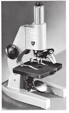
A good microscope is the first and most important piece of equipment for your studies. Of course even the simplest and cheapest “magic tube” will unfold many secrets and new wonders; but everyone, once he has got beyond the simplest beginnings, will feel the need to delve further into the apparently puzzling things he sees, and to make more thorough observations requiring greater magnifications—in short, to see more. The disappointment is great when the microscope no longer “co-operates”—when the limits of its powers have been reached.
A really useful microscope is so made that it can be improved with the purchase of additional optical equipment. A good standard model is not very much more expensive than the widely used, but usually disappointingly small, student’s microscope. It offers the great advantage that by and by, as your purse permits, it can be made into a complete scientific instrument. A simple and therefore inexpensive instrument is sufficient to begin with, but it should be a model that can be elaborated. For ordinary purposes a
in. and a
in. objective and a × 5 eyepiece will suffice, but it is advisable to purchase a condenser at the outset. An instrument which is not too expensive and may later be built up is shown in Fig. 1.
HOW A MICROSCOPE IS CONSTRUCTED
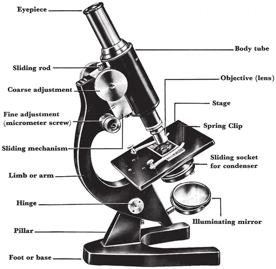
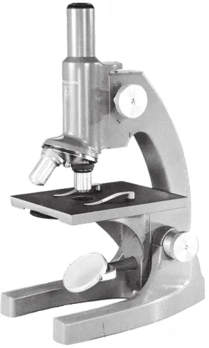
The microscope in Fig. 1 rests on a horseshoe-shaped base or foot from which a low, solid column rises. To this column the upper part of the microscope is joined with a simple, quite tight hinge, which enables the body tube of the microscope to be tilted to an angle of 45°; this permits a comfortable position for the head of the observer. A short distance above the hinge is a roomy, square object stage, on which the slide is firmly held by two spring clips. Above the stage there is a sturdy limb which rises above the center of the stage; this holds the tube that carries the lenses. The objective is screwed to the lower end of the tube, and the upper end holds the eyepiece.
Fig. 1. The parts of the microscope.
For coarse focusing of the microscope there is a rack and pinion adjustment controlled by two large knurled knobs: this moves the tube up and down rapidly. The fine adjustment is made by a second motion controlled by two smaller knobs, the micrometer screws.
With the fine adjustment it is possible to move the tube a minute distance, measured in hundredths of a millimeter. The mechanism for this adjustment is contained within the top of the limb (arm).
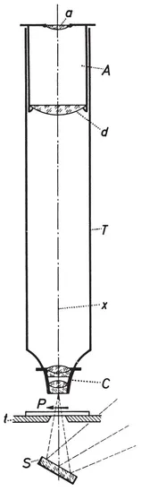
The optical arrangement of the microscope is shown schematically in Fig. 2. You see here a cutaway view through the optical axis of an assembled microscope, represented here by line x which passes through the central point of the lenses. C is the system of lenses in the objective. These lenses are compound or achromatic, and give an image that is without colored edges. Each lens does, in fact, consist of two or three separate lenses made of different kinds of glass.
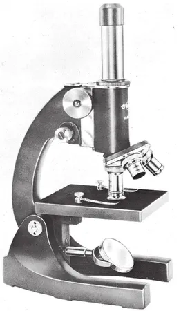
MODELS OF MICROSCOPES
Reichert (Viennese) “Biozet” microscope with micro-drawing attachment
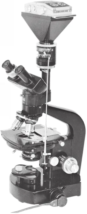
Zeiss-Brinkman universal microscope equipment for fluorescence
Bausch and Lomb teaching microscope
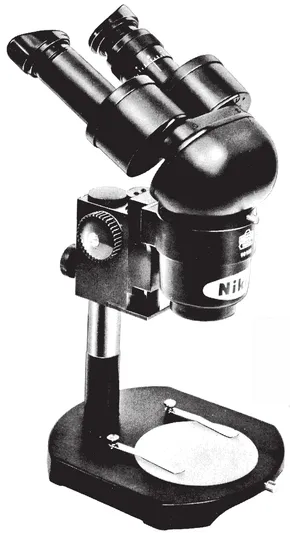
(Above) Nikon (Japanese) stereoscopic microscope with one set of matched objectives
(Below) Wild (German) research microscope with attached 35 mm camera
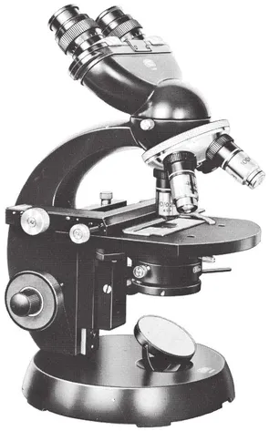
(Left) Carl Zeiss laboratory microscope with inclined binocular tube
(Left, below) American Optical laboratory student microscope
(Below) Swift teaching microscope
Beneath the objective is the stage t, a metal plate with a hole in the center, on which the slide preparation P is placed and lighted by a small beam. The objective C is screwed into the lower end of a metal tube T which is blackened internally. At the upper end of the tube there is a second system of lenses A. It consists of the two lenses a and d, between which there is usually a diaphragm, and is called the ocular or eyepiece, because it is here that the eye (oculus) of the observer is applied. Under the stage there is an illuminating mirror S, which reflects the necessary light through the hole in the stage on to the slide. It follows that the object being examined must be transparent. As Fig. 2 shows, the optical axis x (the tube axis) passes through the exact center of all parts of the instrument. This is absolutely necessary if the microscope is to deliver sharp, undistorted images.
Fig. 2. (left) Cross-section through the optical part of a microscope.
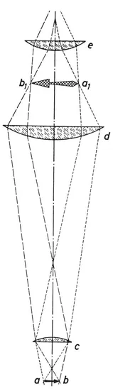
Fig. 3. (right) Schematic diagram of the path of the light rays in the microscope.
How the optical components work is shown in Fig. 3. The objective c is represented by only one lens for the sake of simplicity. It projects an exact and therefore photographable image of an object, which is represented by lines a to b. The ocular lens d breaks up these rays which in the diaphragm behind d are united in an enlarged but reversed image b1 to a1. This magnified image is seen through lens e of the eyepiece as if it were seen through an ordinary magnifying glass; the observer sees a repeated, if only weakly magnified, clear image of the object. Because the common Huygenian eyepiece cannot “erect” the image, the view seen in the microscope is reversed. It follows that if you wish to move the examined object to the right or left, or up or down, you must move the slide in the opposite direction to that desired. This may take a little getting used to.
Like any good microscope, the Humboldt may be equipped with several objective lenses (e.g. 1-in.,
-in. and
-in.) and several eyepieces (e.g. × 5 and X 12). The objective lenses are corrected to suit an exact tube length; in the case of the Humboldt this is 170 mm.1 Objectives and eyepieces can be combined in many ways, so as to give a great range of magnifications (explained in a chart which comes with the instrument). The total magnification of a microscope is determined by a combination of the objective and eyepiece, and is the product of the two magnifications. An objective magnifying 30 diameters, used with an eyepiece of 6 diameters, would give a total magnification of 180 diameters. The magnifying power of the objectives and eyepieces depends upon their focal lengths, and a system magnifies more highly as its focal length decreases.2
At this point it might be well to mention that, contrary to common opinion, strength of magnification does not determine the value of a microscope. Much more important than the power of magnification is a microscope’s so-called “resolution”. This is the power of an optical system to separate minute dots or lines so that details can be distinguished. With a good microscope and the most advantageous lighting the best attainable detail lies around 0.2μ (1μ=1 micron=1/1000 mm.). The tiniest bacteria, which are a little bigger than 0.3μ, can be seen with the help of an exceptional objective—the so-called “oil-immersion lens”. Viruses, on the other hand, which are considerably smaller, cannot be seen with an optical microscope at all.
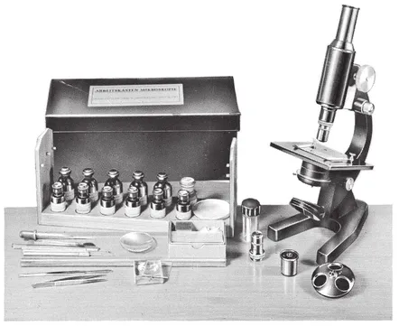
Fig. 4. Microscopy work kit.
The powers of detail of our eyes are much less than that of a usable microscope—and that is why we can see more with the microscope than with the naked eye. The objective lenses ...
Table of contents
Title Page
Copyright Page
Table of Contents
Foreword
1. The Microscope and Essential Tools
2. How to Use the Microscope
3. Examining Simple Preparations
4. Insect Preparations
5. Exploring a Drop of Water
6. The Structure of Plants
7. The Microscopic Structure of Animals
8. Bacteria
9. Microphotography
Appendix - Microtome Technique
Index
Frequently asked questions
Yes, you can cancel anytime from the Subscription tab in your account settings on the Perlego website. Your subscription will stay active until the end of your current billing period. Learn how to cancel your subscription
No, books cannot be downloaded as external files, such as PDFs, for use outside of Perlego. However, you can download books within the Perlego app for offline reading on mobile or tablet. Learn how to download books offline
Perlego offers two plans: Essential and Complete
Essential is ideal for learners and professionals who enjoy exploring a wide range of subjects. Access the Essential Library with 800,000+ trusted titles and best-sellers across business, personal growth, and the humanities. Includes unlimited reading time and Standard Read Aloud voice.
Complete: Perfect for advanced learners and researchers needing full, unrestricted access. Unlock 1.5M+ books across hundreds of subjects, including academic and specialized titles. The Complete Plan also includes advanced features like Premium Read Aloud and Research Assistant.
Both plans are available with monthly, semester, or annual billing cycles.
We are an online textbook subscription service, where you can get access to an entire online library for less than the price of a single book per month. With over 1.5 million books across 990+ topics, we’ve got you covered! Learn about our mission
Look out for the read-aloud symbol on your next book to see if you can listen to it. The read-aloud tool reads text aloud for you, highlighting the text as it is being read. You can pause it, speed it up and slow it down. Learn more about Read Aloud
Yes! You can use the Perlego app on both iOS and Android devices to read anytime, anywhere — even offline. Perfect for commutes or when you’re on the go. Please note we cannot support devices running on iOS 13 and Android 7 or earlier. Learn more about using the app
Yes, you can access The Microscope and How to Use It by Dr. Georg Stehli in PDF and/or ePUB format, as well as other popular books in Tecnologia e ingegneria & Scienze applicate. We have over 1.5 million books available in our catalogue for you to explore.