"Joseph Sheppard has been favorably compared to practically every Renaissance master…he is without peer among modern realists for his ability to impart a warm verisimilitude to the figure." — Artspeak magazine.
In this highly praised guidebook, Joseph Sheppard, a versatile and influential artist and teacher who is widely recognized as a master of figure drawing, introduces an innovative approach to drawing the human form. Beginning by reviewing the basics of anatomy, he makes his principal focus the specifics of surface anatomy.
Rather than depicting in detail the muscles and bone that lie beneath the skin, as do most books on artistic anatomy, this book concentrates on how the position and movement of muscles and bones affect the surface forms of live models. The effects are masterfully demonstrated in over 170 of Joseph Sheppard's own drawings of many different live models in front, back, and side views, and in various standing, sitting, kneeling, crouching, reclining, and twisting poses.
Each drawing is accompanied by two diagrams, one for bones, one for muscles, which specifically show how surface forms are created by the definitive shapes beneath the skin. The superb quality of Joseph Sheppard's drawings, the wide range of poses he illustrates, and the effectiveness of his approach in this book — now in its first paperback edition — will help artists at all levels improve and refine their skills in drawing the living figure.
- 144 pages
- English
- ePUB (mobile friendly)
- Available on iOS & Android
eBook - ePub
Drawing the Living Figure
About this book
Trusted by 375,005 students
Access to over 1.5 million titles for a fair monthly price.
Study more efficiently using our study tools.
Information
Topic
ArtSubtopic
Art TechniquesChapter One
REVIEW of ANATOMY
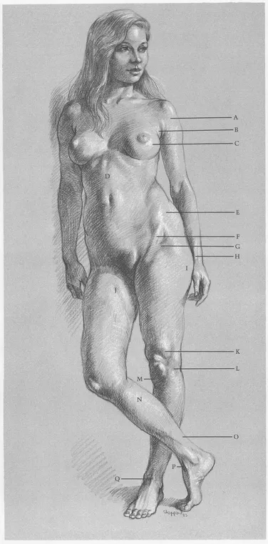
SURFACE ANATOMY
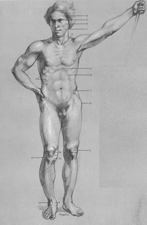
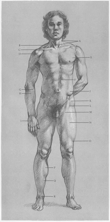
Muscles of skull are thin; bone is close to surface. Skull creates shapes of (A) forehead, (B) cheek, (C) jaw. (D) Eye is egg-shaped, sits in eye socket. (E) Clavicles start at head of (F) sternum, rising upward and back toward outside of shoulder. Note direction of first five ribs attached to sternum: first rib goes upward; second moves straight across; others point downward. (G) End of sternum protrudes. (H) Cavity of rib cage forms arch. (I) Lower ribs slant down from back to front. Pelvis holds stomach like basin. (J) Pelvic crest is prominent. (K) Hipbone is close to skin, clearly seen on male. Note oval shape of (L) kneecap. Silhouette of knee is created by (M) end of femur, (N) head of tibia. (O) Head of fibula creates bump. Ankle is formed by (P) end of tibia, (Q) end of fibula, making hinge joint for foot.
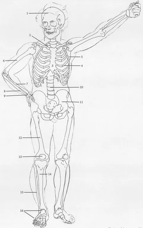
BONES
(1) Skull. (2) Clavicle. (3) Sternum. (4) Rib cage. First seven ribs attach by cartilage to sternum. Each of next three ribs attaches by cartilage to rib above. Eleventh, twelfth ribs do not attach to sternum and are called “floating ribs.” (5) Scapula. (6) Humerus. (7) Radius. (8) Ulna. (9) Wrist. Eight bones of wrist are treated here as one unit. (10) Spinal column is treated here as column of simple discs. (11) Pelvis. (12) Femur. (13) Kneecap. (14) Tibia. (15) Fibula attaches to rear of tibia head, intersecting tibia at ankle and descending further, thus making outside of ankle lower than inside. (16) Bones of foot.
MALE FIGURE, FRONT VIEW
SURFACE ANATOMY
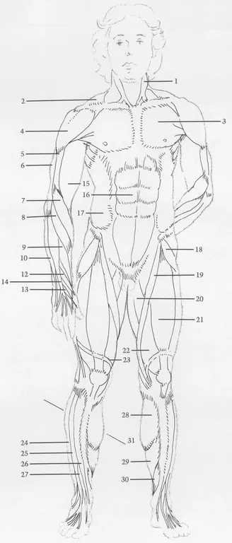
(A) Sternomastoid muscles create “V” shape. (B) Trapezius creates shoulder silhouette. (C) Pectoralis and deltoid meet to form cavity between them. (D) Note division of deltoid. (E) Pectoralis inserts into arm under deltoid. (F) Rectus abdominis is divided. (G) Long supinator and (H) wrist extensor both cross over from outside of elbow to thumb side of wrist. (I) External oblique inserts into top of pelvic crest. (J) Tendons of finger extensor are distinct. (K) Tensor fasciae latae angles toward outer contour. (L) See upside-down “V” where (M) sartorius and tensor fasciae latae overlap (N) rectus femoris. When knee is locked, (O) band of Richer pulls muscles in. (P) Gastrocnemius and (Q) soleus are calf muscles that attach in back of leg; they are seen from front. (R) Tendon of big toe extensor is prominent.
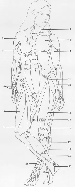
MUSCLES
(1) Stermomastoid. (2) Trapezius originates in back, inserts on top outside area of clavicle. (3) Pectoralis. (4) Deltoid. (5) Brachialis. (6) External head of triceps (one of three heads of triceps muscle). (7) Long supinator (turns forearm, palm out). (8, 9, 10) Extensors of wrist. (11) Abductor of thumb (pulls thumb toward back of hand). (12, 13) Extensors of thumb. (14) Extensor of fingers. (15) Biceps. (16) Rectus abdominis. (17) External oblique. (18) Tensor fasciae latae. (19) Sartorius. (20) Abductors. Several muscles are treated here as one unit. (21) Rectus femoris. (22) Vastus. Two parts are treated as one large muscle under rectus femoris. (23) Band of Richer changes shape of thigh when knee is locked. (Many bands that hold muscles in place are omitted). (24) Long peroneus. (25) Long extensor of toes. (26) Tibialis anterior. (27) Extensor of big toe. (28) Gastrocnemius. (29) Soleus. (30) Long flexor of toes. (31) Inside calf muscle mass is lower than outside mass.
MALE FIGURE, FRONT VIEW
SURFACE ANATOMY
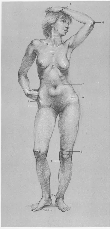
(A) Jawbone determines contour of face. (B) Note slant of clavicle. (C) Sternum shows rib attachments. (D) Cavity of ribcage is narrower on female than on male. (E) Lower ribs are evident. (F) Female pelvis is wider than male. (G) Crest of pelvis is partly covered by body fat. (H) Hipbone is close to skin, creating cavity on hip surface of females and fat males. (I) Kneecap and fat below form figure “8”. (J) Shape of head of tibia slants inward. (K) Inside of ankle is always higher than outside. (L) End of ulna is prominent on little finger side of wrist. (M) Head of ulna forms elbow.
BONES
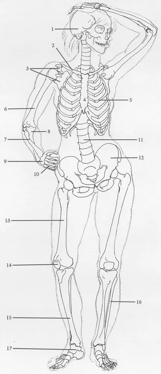
(1) Skull. (2) Clavicle. (3) Scapula. Clavicle and scapula form shoulder socket for humerus—a ball and socket joint. (4) Sternum. (5) Rib cage. (6) Humerus. (7) Radius is always on outside of elbow and thumb side of wrist. Radius head, at elbow, is small. End of radius, at wrist, is large. (8) Ulna is on inside of elbow. Head is large; end is small. (9) Wrist. (10) Bones of the palm. (11) Spinal column. (12) Pelvis. Female pelvis is usually wider than male with crests projecting farther forward. (13) Femur. (14) Kneecap. (15) Tibia. (16) Fibula. Inside of ankle is always higher than outside. (17) Bones of foot.
FEMALE FIGURE, FRONT VIEW
SURFACE ANATOMY
(A) Deltoid attaches to clavicle. (B) Pectoralis lies beneath (C) breast. (D) Vertical division of rectus abdominis is distinct—except on extremely fat figures. (E) External oblique and female body fat cover most of pelvic crest. (F) Sartorius helps to form (G) upside down “V” shaped cavity. (H) Tendon of thumb extensor makes sharp ridge. (I) Female body fat covers hipbone. (J) Indentation is formed by sartorius. (K) Outside of vastus is prominent when knee is locked. (L) Iliotibial band descends outside of thigh like stripe, attaches to outside of tibia head. (M) Small fat deposit appears under kneecap. (N) Calf muscles attach to heel bone by (O) Achilles tendon. (P) Tibialis anterior tendon makes bridge between ankle and foot. (Q) Tendon of toe extensor is prominent.
MUSCLES
(1) Sternomastoid. (2) Deltoid. (3) Pectoralis attaches along sternum and clavicle. (4) Biceps. (5) Brachialis. (6) Rectus abdominis. (7) External oblique. (8) Group of thumb muscles. Three muscles run obliquely across back of forearm into thumb. (9) Rectus femoris. (10) Vastus (inside part). (11) Tensor fasciae latae. (12) Gluteus. (13) Sartorius descends across front of thigh into inside head of tibia. (14) Vastus (outside part). (15) Iliotibial band. (16) Biceps femoris (biceps of leg). (17) Long extensor of toes. (18) Long peroneus. (19) Gastrocnemius. (20) Soleus. (21) Peroneus tertius. (22) Tibialis anterior. (23) Extensor of big toe.
FEMALE FIGURE, FRONT VIEW
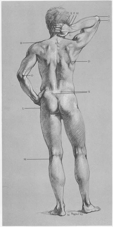
SURFACE ANATOMY
(A) First four vertebrae of ribcage are distinct. (B) Note slight “step down” where head of scapula joins end of clavicle. (C) Edge of scapula protrudes. (D) Lower end of scapula swings upward and outward when arm is raised. (E) Knuckles appear where palm bones join finger bones. (F) Wrist forms flat area between hand and forearm. (G) Radius is longer than (H) ulna at wrist. (I) Hook of ulna ...
Table of contents
- DOVER BOOKS ON ART INSTRUCTION
- Title Page
- Copyright Page
- Dedication
- Table of Contents
- INTRODUCTION
- Chapter One - REVIEW of ANATOMY
- Chapter Two - STANDING FIGURE
- Chapter Three - SEATED FIGURE
- Chapter Four - KNEELING FIGURE
- Chapter Five - CROUCHING FIGURE
- Chapter Six - RECLINING FIGURE
- Chapter Seven - TWISTING FIGURE
- SUGGESTED READING
- INDEX
Frequently asked questions
Yes, you can cancel anytime from the Subscription tab in your account settings on the Perlego website. Your subscription will stay active until the end of your current billing period. Learn how to cancel your subscription
No, books cannot be downloaded as external files, such as PDFs, for use outside of Perlego. However, you can download books within the Perlego app for offline reading on mobile or tablet. Learn how to download books offline
Perlego offers two plans: Essential and Complete
- Essential is ideal for learners and professionals who enjoy exploring a wide range of subjects. Access the Essential Library with 800,000+ trusted titles and best-sellers across business, personal growth, and the humanities. Includes unlimited reading time and Standard Read Aloud voice.
- Complete: Perfect for advanced learners and researchers needing full, unrestricted access. Unlock 1.5M+ books across hundreds of subjects, including academic and specialized titles. The Complete Plan also includes advanced features like Premium Read Aloud and Research Assistant.
We are an online textbook subscription service, where you can get access to an entire online library for less than the price of a single book per month. With over 1.5 million books across 990+ topics, we’ve got you covered! Learn about our mission
Look out for the read-aloud symbol on your next book to see if you can listen to it. The read-aloud tool reads text aloud for you, highlighting the text as it is being read. You can pause it, speed it up and slow it down. Learn more about Read Aloud
Yes! You can use the Perlego app on both iOS and Android devices to read anytime, anywhere — even offline. Perfect for commutes or when you’re on the go.
Please note we cannot support devices running on iOS 13 and Android 7 or earlier. Learn more about using the app
Please note we cannot support devices running on iOS 13 and Android 7 or earlier. Learn more about using the app
Yes, you can access Drawing the Living Figure by Joseph Sheppard in PDF and/or ePUB format, as well as other popular books in Art & Art Techniques. We have over 1.5 million books available in our catalogue for you to explore.