Small Animal Dermatology for Technicians and Nurses
eBook - ePub

Small Animal Dermatology for Technicians and Nurses

  1. English
  2. ePUB (mobile friendly)
  3. Available on iOS & Android
eBook - ePub

Small Animal Dermatology for Technicians and Nurses

About this book

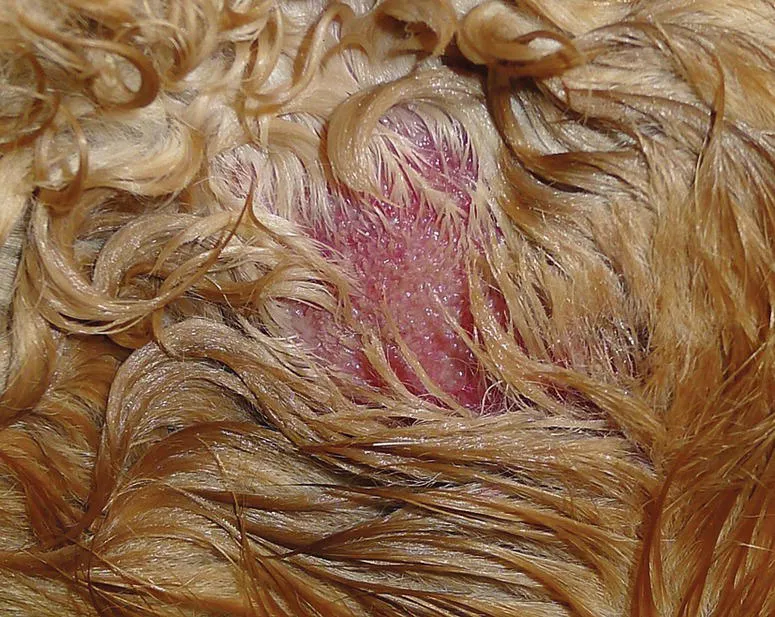
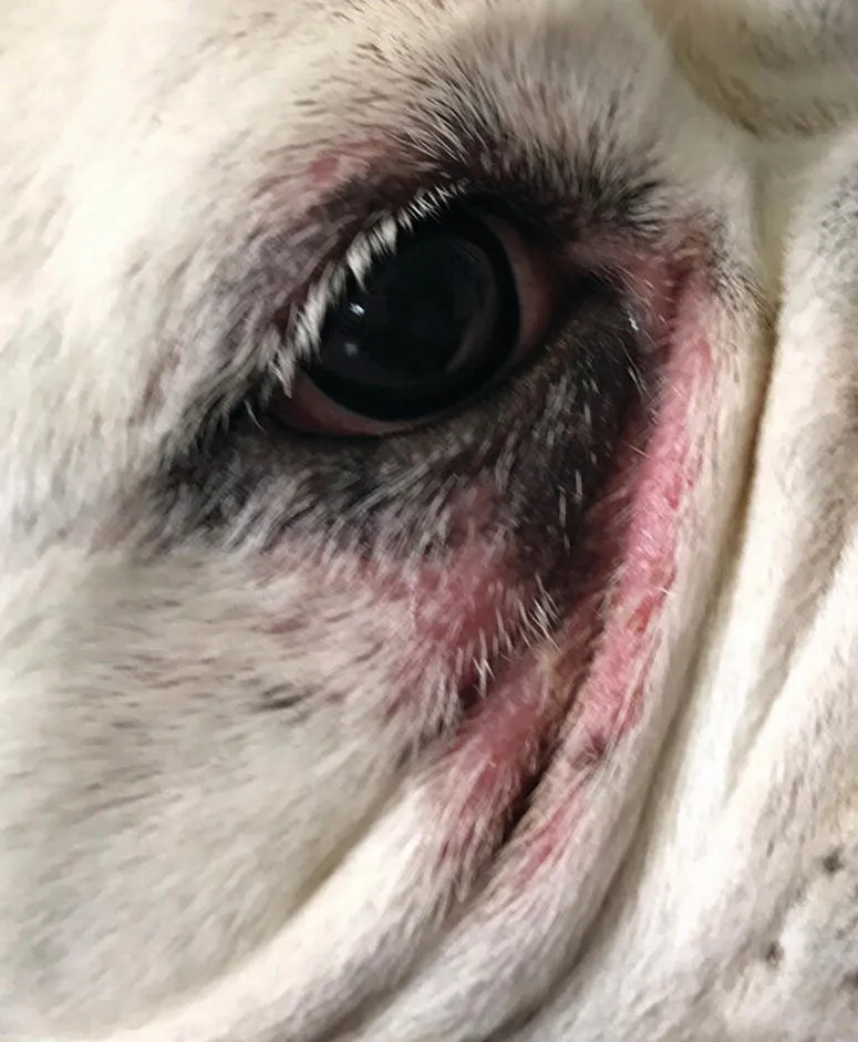
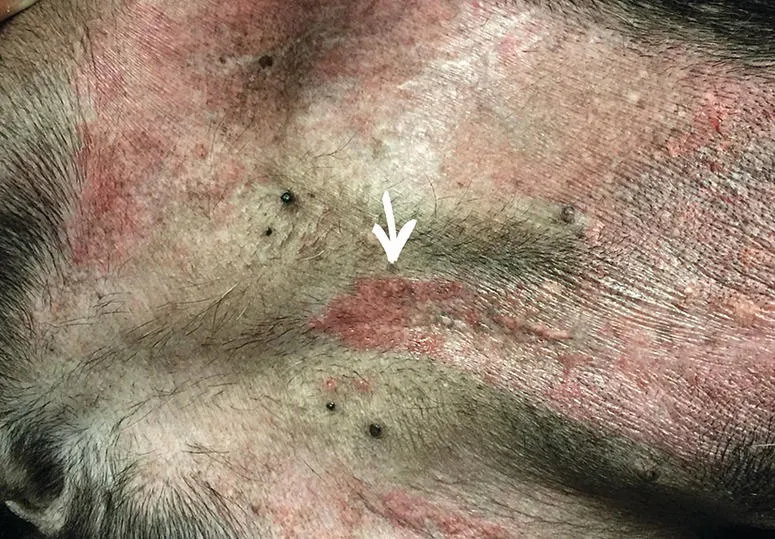
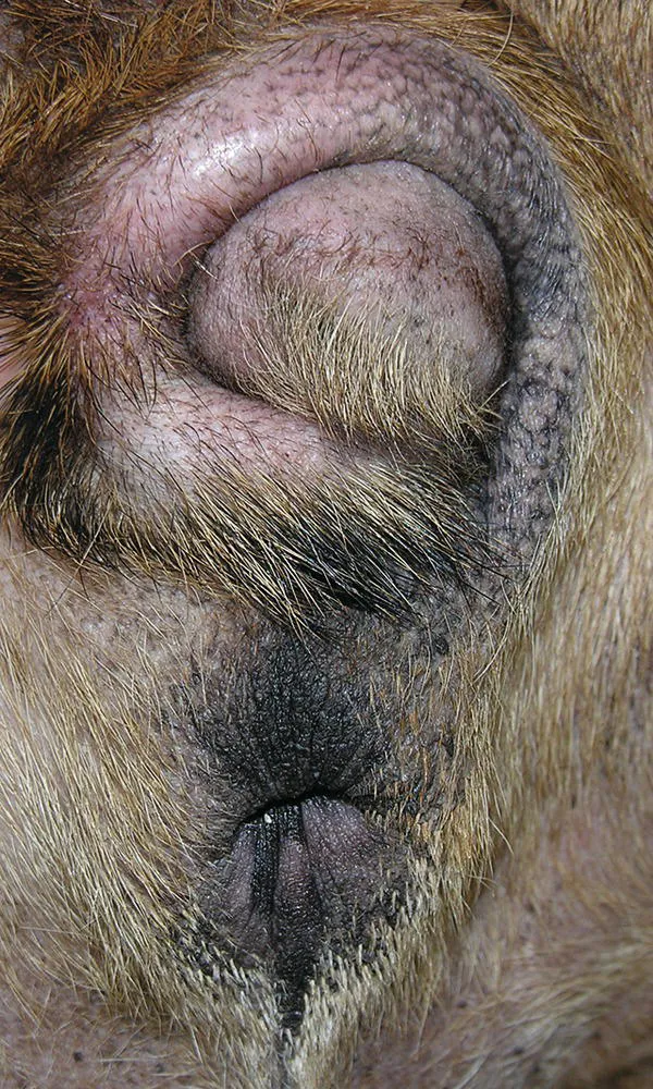
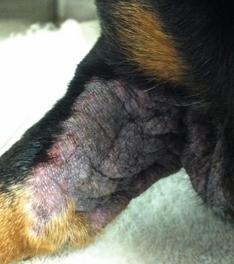
Small Animal Dermatology for Technicians and Nurses is a practical manual specifically designed for veterinary technicians, nurses, and other staff members. This easy-to-read book covers common dermatologic diseases frequently seen in private practice, with information on history taking, sample collection, diagnostic testing, therapeutic options and patient follow up. Each disease described includes important background information to help technicians explain treatment plans and improve client compliance.

The book presents step-by-step instructions with illustrative, full-color photographs to aid in accurately collecting samples and performing in-house diagnostics. The authors have drawn on their real-world experience as practicing dermatologists and dermatology technicians to create a must-have guide for those working in the small animal veterinary field. This important resource:

• Provides practical information on veterinary dermatology tailored to veterinary technicians and nurses

• Offers details and tips on history taking, sample collection, diagnostic testing, and patient follow up

• Takes a practical, easy-to-follow approach with illustrative, full-color photographs that demonstrate techniques

• Supports veterinary staff by providing a solid foundation in dermatology that can improve communication with clients

• Includes all the information technicians need to support their veterinarians in managing dermatology cases with confidence

Written for veterinary technicians, nurses, and veterinary technology and nursing students, Small Animal Dermatology for Technicians and Nurses offers an indispensable reference for any veterinary team member assisting with common small animal dermatology cases.

Frequently asked questions

Yes, you can cancel anytime from the Subscription tab in your account settings on the Perlego website. Your subscription will stay active until the end of your current billing period. Learn how to cancel your subscription.
No, books cannot be downloaded as external files, such as PDFs, for use outside of Perlego. However, you can download books within the Perlego app for offline reading on mobile or tablet. Learn more here.
Perlego offers two plans: Essential and Complete
  • Essential is ideal for learners and professionals who enjoy exploring a wide range of subjects. Access the Essential Library with 800,000+ trusted titles and best-sellers across business, personal growth, and the humanities. Includes unlimited reading time and Standard Read Aloud voice.
  • Complete: Perfect for advanced learners and researchers needing full, unrestricted access. Unlock 1.4M+ books across hundreds of subjects, including academic and specialized titles. The Complete Plan also includes advanced features like Premium Read Aloud and Research Assistant.
Both plans are available with monthly, semester, or annual billing cycles.
We are an online textbook subscription service, where you can get access to an entire online library for less than the price of a single book per month. With over 1 million books across 1000+ topics, we’ve got you covered! Learn more here.
Look out for the read-aloud symbol on your next book to see if you can listen to it. The read-aloud tool reads text aloud for you, highlighting the text as it is being read. You can pause it, speed it up and slow it down. Learn more here.
Yes! You can use the Perlego app on both iOS or Android devices to read anytime, anywhere — even offline. Perfect for commutes or when you’re on the go.
Please note we cannot support devices running on iOS 13 and Android 7 or earlier. Learn more about using the app.
Yes, you can access Small Animal Dermatology for Technicians and Nurses by Kim Horne, Marcia Schwassmann, Dawn Logas, Kim Horne,Marcia Schwassmann,Dawn Logas in PDF and/or ePUB format, as well as other popular books in Medicine & Veterinary Medicine. We have over one million books available in our catalogue for you to explore.

Information

Year
2019
Print ISBN
9780470958155
eBook ISBN
9781119108627
Edition
1

Section II
Skin and Ear Diseases

2
Bacterial Infections

Dawn Logas and Christie Yamazaki Delan

Introduction

Pyodermas are usually classified by the skin layers that are involved. Surface pyodermas affect mainly the stratum corneum, the outermost layer of the epidermis. Superficial pyodermas affect the entire epidermis and the hair follicles. Deep pyodermas also involve the dermis and occasionally the subcutis (Noli and Morris 2012).
Since this chapter is about common bacterial infections seen in practice, we will limit our discussion to infections caused by Staphylococcal bacteria, primarily Staphylococcus pseudintermedius, the Staphylococcus species found most commonly on the dog. Staphylococcus schlieferi and Staphylococcus aureus can also cause canine and feline pyodermas, but are much less common. Detailed information about other bacterial skin infections can be found in references listed in the recommended reading section at the end of this chapter. Canine pyodermas will be discussed in more depth than feline pyodermas, since they are seen much more often in clinical practice.

Pathogenesis

Dogs and cats, like all other mammals, have a variety of bacteria that normally inhabit their skin surface. These bacteria make up the host epidermal microbiome, whose role in skin function and development is still not completely understood. Under normal circumstances these bacteria are kept in check by the host epidermal barrier and the skin's immune system. The presence of another dermatological disease such as allergic dermatitis, endocrinopathy, autoimmune disease, or parasitic infestation can compromise the epidermal barrier and the skin immune system function. This allows these resident bacteria to multiply and become pathogenic, causing the skin disease commonly referred to as pyoderma. For this reason, these infections are considered a secondary problem. Therefore, diagnosis and control of the underlying disease is essential in the management and prevention of these bacterial infections. Client education is consequently extremely important so the owner understands that not only the active infection must be controlled, but the underlying condition as well.

Surface Pyodermas

Surface pyodermas include pyotraumatic moist dermatitis (hot spots) and intertrigo (skin fold dermatitis).

Clinical Features

Pyotraumatic moist dermatitis (Figure 2.1) is caused, as the name suggests, by self‐trauma, usually licking. Therefore, areas of moist dermatitis found in places where the dog cannot lick such as under the ear are not true hot spots and will be discussed later in this chapter. Hot spots are usually extremely pruritic and can come up within a few hours. They are frequently associated with flea bite allergy, so are commonly found on the caudal half of the body. They consist of an erythematous, moist, partially to completely alopecic, exudative area of skin that is pruritic and sometimes painful to the touch.
Image described by caption.
Figure 2.1 Dog with pyotraumatic moist dermatitis on the lateral thigh; note erythema and moist exudate.
Source: Courtesy of Wayne Rosenkrantz DVM, DACVD, Animal Dermatology Clinic, Tustin, CA.
Intertrigo can develop in any dog with skin folds. This condition is common in brachycephalic breeds such as pugs and English bulldogs, but can also be seen in obese dogs of any breed. The lesions can be found anywhere there are skin folds. Some of the more commonly affected areas are the face, ventral neck, axillae, intermammary space, perivulvar fold, and tail fold (Figures 2.22.4). The lesions usually consist of an area of swollen, erythematous skin that is odiferous and moist. In severe cases the skin may be eroded or ulcerated and painful. Pruritus varies from absent to severe.
Close-up of a dog’s eye displaying intertrigo of facial area.
Figure 2.2 Intertrigo of facial fold area.
Source: Courtesy of Amy Weller, Dermatology Clinic for Animals, Tacoma, WA.
Image described by caption.
Figure 2.3 Dog with intertrigo of umbilical fold (white arrow).
Source: Courtesy of Wayne Rosenkrantz DVM, DACVD, Animal Dermatology Clinic, Tustin, CA.
Image described by caption.
Figure 2.4 Dog with intertrigo of tail fold; note the swollen appearance and hyperpigmentation.
Surface pyodermas are also seen in areas of chronic dermatitis where the skin has thickened and lichenified, forming small folds and crevices (Figure 2.5). These areas can also be fairly odiferous with a great deal of exudate. They are usually pruritic.
Image described by caption.
Figure 2.5 Dog with chronic dermatitis showing alopecia, lichenification, and hyperpigmentation in the axilla.
Source: Courtesy of Amy Weller, Dermatology Clinic for Animals, Tacoma, WA.

Diagnostic Tests

Pyotraumatic dermatitis and intertrigo usually present with a classic history and clinical appearance, so are typically diagnosed on physical examination. Cytology should be done to look for the presence of inflammatory cells, bacteria, and yeast. With pyotraumatic moist dermatitis, you would expect to find bacteria on your cytology. Intertrigo can be associated with both bacteria and yeast overgrowths. By identifying the bacteria as cocci (usually Staphylococcus sp.) or rods (Pseudomonas, Proteus, or Escherichia coli), the cytology will help determine which topical therapies will be best to use.
It is also important to look for the underlying cause of the surface bacterial infection. Pyotraumatic de...

Table of contents

  1. Cover
  2. Table of Contents
  3. List of Contributors
  4. Preface
  5. Acknowledgments
  6. Introduction
  7. Section I: The Role of the Veterinary Technician and Nurse
  8. Section II: Skin and Ear Diseases
  9. Section III: Allergic Skin Diseases
  10. Section IV: Parasitic Skin Diseases
  11. Glossary of Terms
  12. Index
  13. End User License Agreement