- 164 pages
- English
- ePUB (mobile friendly)
- Available on iOS & Android
eBook - ePub
Trusted by 375,005 students
Access to over 1.5 million titles for a fair monthly price.
Study more efficiently using our study tools.
Information
Topic
MedicineSubtopic
CardiologyChapter 1
An introduction to electrocardiography
By the end of this chapter you should be able to:
• define a 12-lead electrocardiogram
• identify indications for recording an electrocardiogram
• appreciate the benefit of using a standardised ECG recording approach
• recognise ECG accuracy will always be limited as some events are outside the control of practitioners and current technology.
Since its introduction into clinical practice the electrocardiogram has become one of the most widely available and utilised investigations. ECGs are recorded in a range of settings by practitioners and others involved in fields such as sport and exercise. Despite this there is limited guidance regarding the recording of the electrocardiogram as most textbooks concentrate upon interpretation. Practitioners must be capable of recording accurate ECGs. If skill and experience in electrocardiography is currently not sufficient this can be changed by focusing on and enhancing present practice. The degree to which a practitioner actively engages in continuing professional development and evidence-based practice will be instrumental in determining how quickly these changes occur and ultimately ensure the highest quality healthcare is available for all patients.
What is a 12-lead electrocardiogram?
A 12-lead ECG is a graphical recording obtained by placing ten electrodes on specific positions on the body surface. This provides twelve different views of the electrical activity generated by the myocardium as it depolarises and repolarises to produce the heartbeat. These changes in voltage create a characteristic pattern of positive and negative deflections called waves which can be captured on paper or viewed on a screen. The most frequently seen waves are P, Q, R, S and T; however there are others including F, f, Δ and U waves. Changes in voltage are measured on the vertical axis whilst time is measured on the horizontal axis. This permits the accurate measurement of heart rate, wave durations and timing intervals, wave voltages and the calculation of cardiac axis to be made. Many of these measurements will only be accurate if the electrodes have been placed in precise anatomical positions on the body surface using a standardised methodology. Recording ECGs using a standardised technique permits:
• ECGs recorded at different times, in different settings and by different practitioners to be compared
• changes over time to be monitored
• responses to treatment to be monitored
• accurate comparison with normal reference values to be made and the confident identification of true physiological abnormalities established.
Essentially it is only by using a standardised recording procedure that misdiagnosis due to incorrect electrode placement and/or lead attachment is prevented.
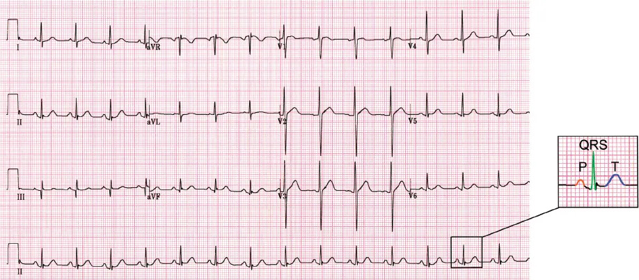
Figure 1.1: Example of a 12-lead ECG highlighting P, QRS complex and T wave.
Indications for recording an electrocardiogram
There are numerous indications for recording an ECG however it is important to understand that the ECG can only directly measure time and voltage; all other information available must be derived or inferred. Research over a number of years has established characteristic patterns and measurements associated with a variety of disease states. However, the sensitivity and specificity of these measurements vary with different pathologies impacting on the ECG’s diagnostic value. In other words, the ECG is useful in some situations such as detecting the presence of a myocardial infarction whilst of limited value in others such as determining the extent of non-viable myocardium. An ECG is indicated for:
• the assessment of patients presenting with chest pain
• unexplained dizziness and/or syncope
• the diagnosis and monitoring of cardiovascular disease
• the assessment of therapy outcomes both beneficial and toxic
• presurgical assessment workup
• risk assessment for cardiac disease in individuals with two or more of the following risk factors: diabetes, hypertension, smoking history, obesity, hypercholesterolaemia, strong family history of cardiac disease
• a family history of sudden death
• the assessment of cardiac effects in coexisting systemic disease e.g. renal failure
• monitoring cardiac transplant success or rejection
• adhering to occupational requirements e.g. airline pilots, divers etc.
• epidemiological studies.
There are no absolute contraindications to recording a resting 12-lead ECG in the clinical environment other than in the uncooperative, abusive patient or a patient that refuses to give consent. However in the field ECGs are typically not recorded where privacy and dignity cannot be protected, safety of the patient or practitioner cannot be guaranteed, or it delays transit time to the detriment of overall patient management. Relative contraindications include trauma and the inability to place electrodes correctly on the chest, extensive skin conditions and burns. Screening the general population is not currently recommended.
How accurate is the ECG?
It is important to recognise that a 12-lead ECG is never 100 per cent accurate. Inaccuracy can arise from non-modifiable patient factors such as respiration patterns, post prandial state (period after a meal), gender, race and body habitus. It may also arise from modifiable practitioner factors such as incorrect electrode placement, lead reversal and poor skin preparation. Adhering to guidelines can help limit the extent of inaccuracy and alert the practitioner to ECG changes they may expect in a specific patient. Even when the ECG is recorded strictly to recommendations, a number of limitations still remain. These include:
• The limited recording period
ECGs provide data over a specific period in time. In A4 format this equates to approximately ten seconds. Anything that occurs before or after this period is not recorded. For example in the patient complaining of frequent palpitations, it is common to record a normal ECG with no arrhythmias and further tests will be required before a diagnosis can be made. On occasion it is possible to infer a past event may have occurred but it is not possible to be confident. For example the presence of Q waves may indicate an old myocardial infarction with non-viable myocardium. But not all Q waves are indicative of necrosis. They are not seen in all patients with myocardial necrosis and when present are more predictive of inferior than anterior non-viable tissue.
• False positives and false negatives
Due to the limited sensitivity and specificity in different disease states, the ECG is prone to producing false positive and false negatives. A false positive occurs when the recorded data indicates a disease or structural change is present when it is not. A false negative suggests that the data is normal in the presence of disease or structural changes. For example a false positive result may occur with ST elevation. In some individuals there is a normal variant called high ST segment take off. This is typically found in young individuals and is not associated with the expected reciprocal ST depression changes or true ischaemic heart disease. The main problem arises when it occurs in the presence of a convincing ischaemic history. Conversely a false negative result may occur in unstable angina. In this case the ECG may be normal yet the patient symptoms represent true ischaemia. Reasons for this include that even with 12 different views of the myocardium there are still electrical blind spots; the expected ST changes can be masked due to pseudonormalisation of the ST/T waves. Pseudonormalisation occurs when abnormal ST/T wave changes revert to a normal pattern. Unless the patient has been monitored or these changes documented at an earlier stage they can be missed. This further highlights the problem that an ECG is a snapshot in time but despite this limitation the ECG remains a cost effective initial diagnostic step in many conditions.
• Reference values and the definition of normality
The majority of ECG reference values regarding timing and voltages are based upon limited patient cohorts from the early days of electrocardiography. We now know that there are gender, ethnic, obesity and normal variations. Consequently inaccuracy in interpretation may occur unless interpretation has been made in line with the correct reference values. Consideration must also be made for normal variants to avoid misinterpretation. This may be a particular problem in relation to the athletic heart. Further problems arise when coexisting pathologies are present. For example caution needs to be taken in diagnosing a myocardial infarction (MI) in the presence of left bundle branch block as the aberrant depolarising pathway conceals the acute changes of MI.
• Day-to-day variability
Consecutive ECGs are prone to natural variation due to the circadian rhythm, changes in physiological state e.g. nervousness or pain, and even after a meal. Heart rate is the most variable factor, followed by the QT interval. ST segment heights are more stable unless electrodes have been placed in different positions. When comparing serial ECGs, minor changes in waveforms and voltages can be expected.
• Inter-/intra-observer variability
ECGs should always be reported by an experienced practitioner. Where computerised interpretation is used the report should be over-read to check for mistakes and omissions. Studies confirm that intra-observer variability in ECG reports (same person reanalysing an ECG) is good. However practitioners do not always agree in their interpretation and inter- observer variability (different individuals analysing the same ECG) is poorer. In theory standardised algorithms applied by the interpretative software packages of ECG machines should overcome the apparent variability. Whilst great improvements in ECG computerised reporting have occurred in recent years, the technology is not fully evolved and will always have to rely on the quality of the data it is fed to analyse.
Although the ECG has many inaccuracies it remains a valuable diagnostic technique. This is because non-modifiable inaccuracies can be accounted for by an experienced practitioner during interpretation and modifiable inaccuracies can be limited by using a standardised ECG recording technique. This allows the ECG to remain an effective and quick diagnostic step in many conditions.
The standardised ECG technique
Papers and textbooks referring to a ‘standard 12-lead ECG’ often mean that a standard set of leads have been recorded rather than the true full meaning which is a standard set of leads recorded from standard electrode positions at a standard calibration setting. Standardisation was first attempted in the 1930s and continues with the most recent recording guidelines produced by the American Heart Association (2007) and the Society of Cardiological Science and Technology (SCST) endorsed by the British Cardiovascular Society (2010). It is important that all those recording ECGs adhere to the same practice standards in relation to the preparation for and recording of an ECG. In this way accuracy, diagnostic quality and confidence in the ECG interpretation can be increased. For standards to be widely accepted:
• they need to be evidence-based and improve practice
• they should be written by eminent personnel in the field of electrocardiography, or by a relevant professional group
• the language used should be accessible to all staff recording ECGs
• they should have been open for a period of consultation and feedback from relevant parties before final publication
• they should be regularly reviewed and updated in the light of new clinical knowledge and technological advances
• they should be made widely available and publicised.
• Put simply recording 12-lead ECGs to an agreed standard is the only way to consistently obtain an ECG of verifiable quality.
At the clinic:
A patient presents with severe chest discomfort and feeling nauseous. Suspecting that it might be of cardiac origin an ECG is requested. In order to obtain this recording urgently no attempt is made to locate the standardised precordial electrode positions or prepare the skin. The recorded ECG shows ST elevation and a decision is made on the basis of symptoms and ECG changes to administer antithrombolytic therapy. Follow-up ECGs recorded in the cardiac unit show no evidence of ST elevation and troponin blood results are normal.
Consider the consequences of failing to follow a standardised guideline for recording the ECG.
Suggest ways in which things could have been done differently, as far as you can justify your opinions and suggestions.
Summary of key points:
• A 12-lead ECG is a graphical representation of the electrical activity generated by the myocardium on depolarisation and repolarisation. It produces characteristic waves with established duration and amplitude limits.
• There are numerous indications for recording an ECG; these range from cardiac presentations, to coexisting pathologies and epidemiological studies.
• Accuracy is limited by non-modifiable and modifiable factors, however whilst accuracy can be improved an ECG can never be 100 per cent accurate.
• Adhering to a standardised recording prot...
Table of contents
- Cover Page
- Title Page
- Copyright
- Contents
- List of Figures
- List of Tables
- Preface
- About the Authors
- Chapter 1 An introduction to electrocardiography
- Chapter 2 Electrocardiographic anatomy
- Chapter 3 Positioning the patient and locating electrode positions
- Chapter 4 Skin preparation
- Chapter 5 ECG electrodes
- Chapter 6 Understanding the lead system and colour codes used for ECG recording
- Chapter 7 Understanding ECG equipment, specification standards and setting choices for recording ECGs
- Chapter 8 Recording and checking the ECG
- Chapter 9 Recognising and reducing artifact in the ECG recording
- Chapter 10 The computer generated report
- Chapter 11 Archiving records
- Chapter 12 Introduction to quality control in ECG recording
- Chapter 13 Infection control in ECG recording
- Chapter 14 Training and continual professional development
- Answers to key review questions
- Index
Frequently asked questions
Yes, you can cancel anytime from the Subscription tab in your account settings on the Perlego website. Your subscription will stay active until the end of your current billing period. Learn how to cancel your subscription
No, books cannot be downloaded as external files, such as PDFs, for use outside of Perlego. However, you can download books within the Perlego app for offline reading on mobile or tablet. Learn how to download books offline
Perlego offers two plans: Essential and Complete
- Essential is ideal for learners and professionals who enjoy exploring a wide range of subjects. Access the Essential Library with 800,000+ trusted titles and best-sellers across business, personal growth, and the humanities. Includes unlimited reading time and Standard Read Aloud voice.
- Complete: Perfect for advanced learners and researchers needing full, unrestricted access. Unlock 1.5M+ books across hundreds of subjects, including academic and specialized titles. The Complete Plan also includes advanced features like Premium Read Aloud and Research Assistant.
We are an online textbook subscription service, where you can get access to an entire online library for less than the price of a single book per month. With over 1.5 million books across 990+ topics, we’ve got you covered! Learn about our mission
Look out for the read-aloud symbol on your next book to see if you can listen to it. The read-aloud tool reads text aloud for you, highlighting the text as it is being read. You can pause it, speed it up and slow it down. Learn more about Read Aloud
Yes! You can use the Perlego app on both iOS and Android devices to read anytime, anywhere — even offline. Perfect for commutes or when you’re on the go.
Please note we cannot support devices running on iOS 13 and Android 7 or earlier. Learn more about using the app
Please note we cannot support devices running on iOS 13 and Android 7 or earlier. Learn more about using the app
Yes, you can access Practical Aspects of ECG Recording by in PDF and/or ePUB format, as well as other popular books in Medicine & Cardiology. We have over 1.5 million books available in our catalogue for you to explore.