Many books on ECG interpretation use simulated ECG tracings. Most of the traces that you find in this book are from real people and of the quality that you will be expected to interpret from in practice.There is now a chapter on Wolff Parkinson White syndrome in this fourth edition, which in addition to the chapters on Hemiblocks, Bi Fascicular and Tri Fascicular Blocks and Paced Rhythms in the last edition make The ECG Workbook a very comprehensive resource for healthcare professionals and students.All the additional chapters adhere to the principles followed set out in the first editions: that the text should be accessible and relevant to all practitioners, regardless of their experience and that the text should always be supported with relevant exercises to reinforce learning.
- English
- ePUB (mobile friendly)
- Available on iOS & Android
eBook - ePub
About this book
Trusted by 375,005 students
Access to over 1.5 million titles for a fair monthly price.
Study more efficiently using our study tools.
Information
Topic
MedicineSubtopic
CardiologyChapter 1
Recording a readable electrocardiogram (ECG)
An ECG is a graphic tracing of electrical patterns produced by the heart. This test is frequently used for patients who have heart problems and is an important diagnostic procedure. However, the ECG has limitations and so it is important to evaluate it in conjunction with the patient’s clinical status. ECG abnormalities can occur in healthy individuals; and it can also be possible for a person to have a heart attack and yet have a normal ECG. The nature of the abnormality and its effect on the patient influence the clinical importance of the findings, so the ECG should never be used in isolation.
Before we start to interpret the ECG, it is important to learn how to obtain a readable recording (see fig 1.1). We will learn in the following chapters that slight changes can have huge implications for the patient. These days it is common for ancillary staff to take on the task of recording ECGs. To the untrained eye a recording may seem readable but it is not until we learn to interpret an ECG recording that we really gain an understanding of the importance of producing a readable tracing. It is possible to misdiagnose patients or miss their diagnosis if the recording is not clear.
Before interpreting the ECG, it is therefore essential to ensure that the recording was obtained correctly. Common errors are incorrect paper speed and standardisation, artefact and incorrect lead placement. Any of these problems can make it extremely difficult, and in some cases impossible, to measure the intervals and the segments that we are going to learn about in this book.
Paper speed and standardisation
The ECG is made up of a series of horizontal and vertical lines that measure the duration and amplitude of the various deflections. The small boxes on the paper are 1 millimetre (mm) in height and I mm in width. The amplitude of the ECG deflection is measured vertically and the duration of the ECG event horizontally. Recordings are usually made at 25 mm per second (mm/s). It is therefore important to ensure that the ECG machine has not been set, at say, 50mm/s before the ECG is recorded. The paper speed should be printed on the ECG itself when it is recorded (see fig 1.2).
A standard deflection (a box that looks like half a rectangle) should be inscribed at the beginning or end of the ECG. The ECG is usually standardised so that the amplitude of a 1 millivolt impulse causes a deflection of 10 mm (see fig 1.3). An increased amplitude (or voltage) usually indicates increased muscle mass of the heart.
Note: If the ECG is not set at 25 mm/s, all the usual ECG measurements that we are about to learn will not apply.

Figure 1.1: A 12 lead ECG with many normal features
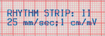
Figure 1.2: The paper (or ‘sweep’) speed printed on an ECG.
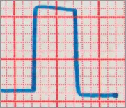
Figure 1.3: The standard deflection at the end of an ECG.
Artefact
To obtain a good-quality ECG tracing you need to make sure that there is no outside interference, as this can create artefact. The three most common causes of artefact are:
1 mains interference
2 patient movement
3 wandering baseline.
Mains interference
Mains interference may produce a fuzzy trace. Too much or too little heat stimulus will produce a tracing that is too thick or too faint. Mains interference usually comes from electrical interaction at or near the patient’s bedside. For this reason, any pumps or electric fans situated nearby should be switched off or left to run on battery while the ECG is being recorded. Interference can also occur if the patient is in contact with metal, such as the end of the bed, or if an ECG lead is in contact with metal (e.g. touching a watch) or the ECG leads are tangled. Recording the ECG with the machine on battery status instead of mains also helps to eliminate mains interference.
Patient movement
If the patient is tense or moving during the recording, artefact will result. What may be a routine procedure to a healthcare professional is not always a routine procedure to a patient. Learning that the healthcare professional wants to take a tracing of their heart is not exactly conducive to relaxation! For instance, patients have been known to express concern that they might get electrocuted if the healthcare professional gets the leads in the wrong order! For all these reasons it is important to explain to the patient the aim of the procedure, that it will not hurt, that it will only take a few minutes and that it will help if they can relax as much as possible, as this will produce a clearer recording. It is often helpful to ask patients to close their eyes and imagine themselves somewhere relaxing.
Many patients feel embarrassed about having their chests exposed for an ECG recording. Always ensure their privacy during the recording. Remember also that some patients may concentrate on your facial expression in an attempt to assess your reaction to the ECG as it is being printed. You should therefore try to keep your expression as neutral as possible.
Wandering baseline
Wandering baseline makes it difficult to identify ECG changes, as many of these changes are measured from the baseline. This problem is often caused by poor electrode contact with the skin. You might need to ask permission to shave some of the hair off the patient’s chest in order to obtain good electrode contact. It may also be necessary to dry the skin if the patient is sweating, or clean the skin if talcum powder has been applied. Ensure that the skin is completely dry after cleaning.
Lead placement
ECG electrodes must be placed in the correct positions on the body. If they are not, changes could appear on the recording that are simply caused by looking at the heart from a slightly different angle. This could easily lead to misdiagnosis. It would also make comparison of the patient’s ECG recordings unreliable.
The limb leads are labelled: R (right), L (left), F (foot) and N (neutral). The R lead should be attached to the patient’s inner, right wrist; the L lead to the inner, left wrist; the F lead to the inner left leg (just above the ankle); and the N lead in the same position on the right leg (see fig 1.4).
Ideally electrodes should be placed over fleshy surfaces, as flesh conducts electricity much better than bone. It is important to have the leads the right way round, otherwise this could change the polarity of the ECG complexes. What is being measured from these leads is simply the difference in electrical potential between two points, so if these points vary slightly (e.g. in a patient with an amputation), one would simply attach the leads higher up the leg.
It is vital, however, to get the position of the chest electrodes correct (see fig 1.5):
1 V1 should be positioned in the fourth intercostal space counting down from the patient’s right sternal notch on the right sternal edge;
2 V2 should be positioned in the fourth intercostal space counting down from the patient’s left sternal notch on the left sternal ...
Table of contents
- Cover Page
- Title Page
- Copyright
- Contents
- Preface to the fourth edition
- About the authors
- 1 Recording a readable electrocardiogram (ECG)
- 2 The electrical conducting system of the heart
- 3 A systematic approach to rhythm strip analysis
- 4 Heart blocks
- 5 Common arrhythmias
- 6 Ectopics and extrasystoles
- 7 The 12 lead ECG
- 8 Axis deviation
- 9 Ischaemia, injury and necrosis
- 10 Sites of infarction
- 11 Bundle branch blocks
- 12 Chamber enlargement
- 13 Hemiblocks, bifascicular blocks and trifascicular blocks
- 14 Paced rhythms
- 15 Wolff-Parkinson-White syndrome
- 16 A systematic approach to ECG interpretation
- Answers to activities
- References
- Glossary
- Index
Frequently asked questions
Yes, you can cancel anytime from the Subscription tab in your account settings on the Perlego website. Your subscription will stay active until the end of your current billing period. Learn how to cancel your subscription
No, books cannot be downloaded as external files, such as PDFs, for use outside of Perlego. However, you can download books within the Perlego app for offline reading on mobile or tablet. Learn how to download books offline
Perlego offers two plans: Essential and Complete
- Essential is ideal for learners and professionals who enjoy exploring a wide range of subjects. Access the Essential Library with 800,000+ trusted titles and best-sellers across business, personal growth, and the humanities. Includes unlimited reading time and Standard Read Aloud voice.
- Complete: Perfect for advanced learners and researchers needing full, unrestricted access. Unlock 1.5M+ books across hundreds of subjects, including academic and specialized titles. The Complete Plan also includes advanced features like Premium Read Aloud and Research Assistant.
We are an online textbook subscription service, where you can get access to an entire online library for less than the price of a single book per month. With over 1.5 million books across 990+ topics, we’ve got you covered! Learn about our mission
Look out for the read-aloud symbol on your next book to see if you can listen to it. The read-aloud tool reads text aloud for you, highlighting the text as it is being read. You can pause it, speed it up and slow it down. Learn more about Read Aloud
Yes! You can use the Perlego app on both iOS and Android devices to read anytime, anywhere — even offline. Perfect for commutes or when you’re on the go.
Please note we cannot support devices running on iOS 13 and Android 7 or earlier. Learn more about using the app
Please note we cannot support devices running on iOS 13 and Android 7 or earlier. Learn more about using the app
Yes, you can access The ECG Workbook by Angela Rowlands, Andrew Sargent in PDF and/or ePUB format, as well as other popular books in Medicine & Cardiology. We have over 1.5 million books available in our catalogue for you to explore.