![]()
Part I
General Information
![]()
CHAPTER 1
Extracellular Matrix Scaffolds for Tissue Engineering and Biological Research
Takashi Hoshiba*a ,b and Tetsuji Yamaoka*c
a Biotechnology Group, Tokyo Metropolitan Industrial Technology Research Institute, 2-4-10 Aomi, Koto-ku, Tokyo 135-0064, Japan,
b National Institute for Materials Science, 1-1 Namiki, Tsukuba, Ibaraki 305-0044, Japan
c Department of Biomedical Engineering, National Cerebral and Cardiovascular Center Research Institute, 6-1 Kishibe Shinmachi, Suita, Osaka 564-8565, Japan,
*E-mail:
[email protected],
[email protected] Cell manipulation is one of the major concerns in medicine, pharmaceuticals, and tissue engineering. Since the extracellular matrix (ECM) plays a pivotal role in regulating cell behavior and function, as well as various soluble bioactive substrates, in vitro reconstitution of ECM with intrinsic functions has been in great demand for medical, pharmaceutical, and tissue engineering applications. However, reconstitution is not easy by conventional chemical and physical methods due to the compositional and structural complexity of the ECM. For this reason, the decellularization technique is increasingly focused upon. In this chapter, we summarize the structures, compositions and functions of the ECM. Additionally, trials mimicking the ECM will be briefly addressed.
1.1 Introduction
The regulation of cell functions is still one of the major challenges for the medical, pharmaceutical, and tissue engineering fields. There are many efforts to regulate cell functions; for example, genetic modification to alter intracellular signaling 1 and stimulation with soluble factors, including growth factors and hormones. 2,3 Matrices with wide diversity being adjacent to cells are also important for regulating cell functions, which are useful as cell regulating cues in a variety of fields. Many scaffolds have been proposed so far, such as metallic, 4 inorganic, 5 and synthetic and natural polymeric ones. 6 These scaffolds regulated the cell functions to some extent. However, the function of these scaffolds was not as good as that of the native extracellular microenvironments. In the in vivo extracellular microenvironments, the cells are supported by the extracellular matrix (ECM) supplying the substrates. The ECM also regulates many cell functions through various modes. Thus, it is a major challenge to develop substrates that mimic the native ECM. Decellularized ECM (dECM), prepared by a technique for the specific removal of cellular components (the decellularization technique), is a promising approach for preparing substrates that mimic the native ECM. In this chapter, we summarize the roles of the ECM in the regulation of cell functions, and we also introduce trials to prepare substrates that mimic the native ECM for medical, pharmaceutical, and tissue engineering applications. The substrates mimicking the native ECM can also be used for comprehensive research into the roles of ECM as well as the above applications.
1.2 General ECM Information
1.2.1 Composition
The ECM is composed of various proteins and carbohydrates. The number of ECM protein types is speculated to be approximately 300. 7 These ECM proteins are categorized into four types: collagens, elastin, proteoglycans, and glycoproteins. 8 Collagens provide the structural strength of the ECM in the strong fibers of tendons, the organic parts of bones and cartilage, the basement membranes, the viscous matrix of the vitreous humor, the dermis, and the capsules around organs. 8 Elastin is one of the major structural proteins found in the native ECM. It determines the elasticity of connective tissues and various organs, including vascular walls, ligaments, skin, and so on. 9,10 Elastin is chemically inert and highly insoluble and possess many covalently cross-linked structures, which are formed between the lysyl residues and make the soluble tropoelastin into insoluble elastin. 10 In addition, elastin was known to be the origin of matrikines, peptides originating from the fragmentation of matrix proteins and presenting biological activities, as described later (see Section 1.2.3.4) 11,12 Proteoglycans are composed of two parts: core proteins and glycosaminoglycans (GAGs). Additionally, free GAGs, such as hyaluronan, can exist in the ECM. GAGs are repeating polymers of disaccharides with carboxyl and sulfate groups; thus, proteoglycans with GAGs can have high negative charges. Due to these negative charges, GAGs chains can be extended and they contain a high water content, leading to space-filling and lubricating functions. 8 Additionally, GAGs can bind to many growth factors to store them in the ECM and to regulate their availability and activity, as explained below. 13,14
In addition to these collagens, elastins, and proteoglycans, there are approximately 200 types of glycoproteins, such as fibronectin and laminin. 8 Many of these glycoproteins can interact with the cells to regulate their functions as addressed in detail below.
The ECM is composed of these molecules with many variations. Moreover, the composition of the ECM differs among different tissues and organs. 15 For example, type I collagen exists in cancellous bone but not in articular cartilage. Instead of type I collagen, type II collagen exists in articular cartilage. Additionally, the composition of the ECM changes according to developmental stages and pathological states to precisely regulate cell functions. 16,19 Due to this compositional and structural complexity, it is difficult to prepare substrates mimicking the native ECM by conventional chemical and physical methods.
1.2.2 Structures
The ECM molecules described above can be assembled to form special structures. Several ECM molecules can interact with the same and/or other ECM molecules to form ECM structures. 20,22 For the cells in epithelial tissues, the ECM molecules are assembled into a sheet form called the basement membrane. 23 There are four fundamental components in the basement membrane: type IV collagen, laminins, nidogen, and perlecan. 23 Both type IV collagen and laminin form their own mesh structures, and these two mesh structures are bound via nidogen. 24 Other basement membrane molecules, including perlecan, are in this type IV collagen–laminin double mesh structure. Similar to epithelial tissues, the cells in muscles, nerves, and blood vessels are also supported by the basement membrane. In stromal tissues, the cells are surrounded by the ECM, which is a gel-like structure. This stromal ECM is composed of molecules including type I collagen, fibronectin, and elastin.
1.2.3 Functions
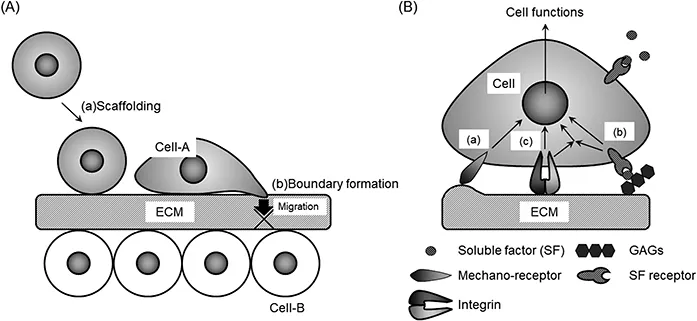
Due to the above compositional complexity of the ECM, the ECM has various functions related to maintaining the homeostasis of living beings. The functions are classified into five categories: (1) scaffolding to maintain tissue and organ structures [Figure 1.1A, label (a)] (2) forming boundaries between the different tissues and organs [Figure 1.1A, label (b)], (3) transducing mechanical signals [Figure 1.1B, label (a)], (4) regulating the activity of soluble factors [Figure 1.1B, label (b)], and (5) directing signal transduction via interaction with the cells [Figure 1.1B, label (c)].
Figure 1.1 The regulatory modes of cell functions by the ECM. (A) Physical roles of ECM. (a) Scaffolding and (b) boundary formation between different tissues. (B) Three modes for the regulation of cell functions. (a) Mechanical signal transduction from the substrates, (b) Signal activation from soluble factors bound to the ECM. (c) Intracellular signal activation by direct interaction with ECM molecules via integrin. GAGs, glycosaminoglycans.
1.2.3.1 Scaffolding to Maintain Tissue and Organ Structures
Scaffolding is the most fundamental role of the ECM. Cells can adhere to many ECM molecules, such as fibronectin, vitronectin, laminin, elastin, and collagen, via various receptors on the cell surface. 25,27 The cells adhering to the ECM molecules can be fixed onto the ECM scaffolds with tissue- or organ-specific structures. For example, epidermal keratinocytes are on the basement membrane of the skin. 23 When the skin is exposed...