Digital models based on data from medical images have recently become widespread in the field of biomechanics. This book summarizes medical imaging techniques and processing procedures, both of which are necessary for creating bone models with finite element methods. Chapter 1 introduces the main principles and the application of the most commonly used medical imaging techniques. Chapter 2 describes the major methods and steps of medical image analysis and processing. Chapter 3 presents a brief review of recent studies on reconstructed finite element bone models, based on medical images. Finally, Chapter 4 reveals the digital results obtained for the main bone sites that have been targeted by finite element modeling in recent years.
Trusted by 375,005 students
Access to over 1.5 million titles for a fair monthly price.
The field of medical imaging has experienced revolutionary progress, with improved accuracy and reduced invasiveness. Medical imaging techniques make it possible to better understand human behavior and are essentially composed of an energy-emitting source able to penetrate the human body. During its penetration, this energy can be absorbed or attenuated at different levels depending on the tissue density and the penetrated atomic number. This process generates signals that can be detected by special detectors specific to the energy source. Mathematical models are then used to manipulate these signals in order to create medical images.
An imaging modality is a specific imaging technique or system used to visualize the inside of the body. According to the used energy source, several modalities can be distinguished. Diagnostic radiology is based on the use of the electromagnetic spectrum beyond the visible light region, such as X-rays used in mammography and computed tomography, whereas magnetic resonance imaging is based on the use of radiofrequencies and ultrasound imaging is based on the use of mechanical energy in the form of high-frequency sound waves.
During the acquisition of a medical image, the acquisition conditions and the technical quality of the image are the two determinant factors of its diagnostic utility. In general, the image quality involves compromises; it improves when the dose of X-rays increases in radiography and computed tomography, when the image acquisition time increases in MRI and when the power levels increase in ultrasound imaging. However, the safety and the comfort of the patient are crucial parameters that should be taken into account during the acquisition process, and an excessive radiation dose should not be applied in pursuit of a perfect image. Indeed, the quality of the image and the safety of the patient must be balanced.
This chapter briefly addresses the four medical imaging modalities that are most commonly used to create FE models of different parts of the human skeleton: X-ray imaging, computed tomography (CT), magnetic resonance imaging (MRI) and ultrasound imaging.
1.2. X-ray imaging
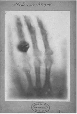
X-ray imaging is the foremost technique that is used to perform medical imaging. The X-rays used in radiography were discovered in 1895 by physicist Wilhelm Roentgen, who created the first radiographic images of the human anatomy (Figure 1.1) (Bushberg and Boone 2011).
Hence, X-ray imaging has gone on to define the radiology field, leading to the emergence of radiologists and specialists in interpreting medical images (Bushberg and Boone 2011).
1.2.1. Definition of X-rays
X-rays fall within the electromagnetic rays (Figure 1.2) that transport radiating energies through space by waves and photons. These rays can be represented by a photon or a wave model and can be categorized according to their energy, frequency fp or wavelength λp (Berger et al. 2018):
[1.1]
where c0 denotes the wave propagation speed, i.e. the speed of light. The photon energy Ep (eV) is directly linked to λp or to fp as follows (Berger et al. 2018):
[1.2]
where һ is Planck’s constant (≈ 6,626069 × 10 Js) and с is the speed of light (≈ 2,99792 × 10s m s−1).
By passing through different materials, X-rays lose a certain amount of energy according to the absorption behavior of each material, which describes the basic principle of traditional X-ray radiography that measures the amount of lost energy. The contrast in the image comes from the difference in the energy amount lost between the different materials. The absorbed energy amount also depends on the dose released during acquisition (Berger et al. 2018).
Figure 1.1.This famous image represents the oldest existing human X-ray image. It was taken on December 22, 1895 by Roentgen, and shows the hand of his wife. This X-ray, clearly showing the bones of the hand as well as two rings on one of the fingers, represents the beginning of diagnostic radiology. Within several months, Roentgen was able to determine t...
Table of contents
Cover
Table of Contents
Introduction
1 Main Medical Imaging Techniques
2 Medical Image Analysis and Processing
3 Recent Methods of Constructing Finite Element Models Based on Medical Images
4 Main Bone Sites Modeled Using the Finite Element Method
Conclusion
References
Index
End User License Agreement
Frequently asked questions
Yes, you can cancel anytime from the Subscription tab in your account settings on the Perlego website. Your subscription will stay active until the end of your current billing period. Learn how to cancel your subscription
No, books cannot be downloaded as external files, such as PDFs, for use outside of Perlego. However, you can download books within the Perlego app for offline reading on mobile or tablet. Learn how to download books offline
Perlego offers two plans: Essential and Complete
Essential is ideal for learners and professionals who enjoy exploring a wide range of subjects. Access the Essential Library with 800,000+ trusted titles and best-sellers across business, personal growth, and the humanities. Includes unlimited reading time and Standard Read Aloud voice.
Complete: Perfect for advanced learners and researchers needing full, unrestricted access. Unlock 1.5M+ books across hundreds of subjects, including academic and specialized titles. The Complete Plan also includes advanced features like Premium Read Aloud and Research Assistant.
Both plans are available with monthly, semester, or annual billing cycles.
We are an online textbook subscription service, where you can get access to an entire online library for less than the price of a single book per month. With over 1.5 million books across 990+ topics, we’ve got you covered! Learn about our mission
Look out for the read-aloud symbol on your next book to see if you can listen to it. The read-aloud tool reads text aloud for you, highlighting the text as it is being read. You can pause it, speed it up and slow it down. Learn more about Read Aloud
Yes! You can use the Perlego app on both iOS and Android devices to read anytime, anywhere — even offline. Perfect for commutes or when you’re on the go. Please note we cannot support devices running on iOS 13 and Android 7 or earlier. Learn more about using the app
Yes, you can access Finite Element Method and Medical Imaging Techniques in Bone Biomechanics by Rabeb Ben Kahla,Abdelwahed Barkaoui,Tarek Merzouki in PDF and/or ePUB format, as well as other popular books in Biological Sciences & Biotechnology. We have over 1.5 million books available in our catalogue for you to explore.