- 232 pages
- English
- ePUB (mobile friendly)
- Available on iOS & Android
eBook - ePub
The Aortic Valve
About this book
This book provides information on the aortic valve. Written in a comprehensive style, it emphasizes the principles behind the development of artificial valves. It covers the principles of valve geometry, tissue structure and function relationships, valve dynamics, fluid dynamics, mechanical stresses, echocardiographic images, mechanisms of valve sounds, valvular pathology, and design and performance of bioprosthetic valves. It enhances our understanding of angiographic and echocardiographic images and calcific stenosis, and will be of value in the development of better prostheses. The Aortic Valve is the ideal text for biomedical engineers and a unique resource for teaching interdisciplinary approaches to medical and engineering students. This work is also an indispensible source for cardiac surgeons, pathologists, cardiologists, and manufacturers of prosthetic valves.
Trusted by 375,005 students
Access to over 1.5 million titles for a fair monthly price.
Study more efficiently using our study tools.
Information
Topic
MedicineSubtopic
CardiologyChapter 1
GEOMETRY OF THE AORTIC VALVE
I. INTRODUCTION
The aortic valve consists of three membranous leaflets and aortic sinuses. The valve is located between the left ventricle and the aorta and its function is to allow the blood to flow in one direction, from the ventricle to the aorta. The valve separates the ventricle from the aorta. The geometry of the valve will be described in four sections: (1) heart and heart valves, (2) valve anatomy, (3) dimensions of the valve, and (4) principles of valve design.
II. HEART AND HEART VALVES
The heart has four chambers (right atrium, right ventricle, left atrium, and left ventricle) and four valves (tricuspid, pulmonary, mitral, and aortic valves) (Figure 1). The tricuspid valve is located between the right atrium and the right ventricle, the pulmonary valve between the right ventricle and the pulmonary artery, the mitral valve between the left atrium and the left ventricle, and the aortic valve between the left ventricle and the aorta. The tricuspid and mitral valves are called atrioventricular valves since they are between the atrium and the ventricle, and the pulmonary and aortic valves are called arterioventricular valves since they are between the artery and the ventricle. The aortic and pulmonary valves are also called semilunar valves because their leaflets have the shape of a half moon. Atrioventricular valves are attached to the heart muscle (myocardium) by means of papillary muscles and fibrous cords and are considered to be active structures responding to myocardial contractions. Semilunar valves, on the other hand, do not have direct attachment of the mobile part of the leaflet to the myocardium, and therefore have been considered in the past to function passively in response to blood flow. It will be shown that parts of the aortic valve are active.
All of the valves permit blood flow in one direction (Figure 1). Oxygen-depleted blood returns from the body via venae cavae to the right atrium and through the tricuspid valve to the right ventricle. It then goes through the pulmonary valve to the pulmonary artery, and to the lungs. Oxygenated blood from the lungs returns via pulmonary veins to the left atrium and through the mitral valve to the left ventricle. It then goes through the aortic valve to the aorta, and finally to the whole body. The flow of blood is achieved by the pumping action of the heart. During ventricular ejection, aortic and pulmonary valves remain open and mitral and tricuspid valves remain closed, and during ventricular filling, aortic and pulmonary valves remain closed and mitral and tricuspid valves remain open.
The aortic valve opens to allow blood to flow into the aorta, and closes to prevent backflow into the ventricle. The valve opens and closes approximately 103, 000 times each day and approximately 3.7 billion times in its life span. This opening and closing of the aortic valve is achieved by the movement of its three leaflets. To create a mental picture of the aortic valve in action, imagine three leaflets opening and closing, like the shutter of a camera, with each heartbeat. Although the aortic valve performs the same task as man-made valves, it is unique in its accomplishments since no man-made valve, to date, can serve that function with the same efficiency and durability. This fact becomes consequential when a diseased aortic valve has to be replaced with a prosthetic device which must now perform the same task (see Chapter 9).
Although diseases of any of the heart valves can compromise health, diseases of the aortic or the mitral valve result in more dire consequences, which therefore puts greater importance on the function of these two valves.
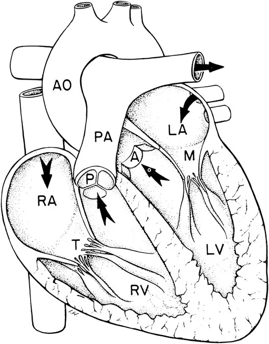
FIGURE 1. Drawing of the heart showing its chambers and valves. RA — right atrium, RV — right ventricle, LA — left atrium, LV — left ventricle, T — tricuspid valve, P — pulmonary valve, M — mitral valve, A — aortic valve, PA — pulmonary artery, AO — aorta. Arrows indicate the path of blood flow.
III. VALVE ANATOMY
The aortic valve consists of three leaflets and three sinuses (Figures 2 and 3). The leaflets are the most mobile parts of the valve and the sinuses are cavities behind the leaflets. At the lower margin, the sinuses become continuous with the left ventricle, and at the upper margin they become part of the ascending aorta. The sinuses represent dilations of the base of the aorta.
When the aortic root is excised and fixed at diastolic pressure with the leaflets closed, its outside wall is seen to be formed by three aortic sinuses ballooning outward in the shape of ellipsoids (Figure 3I). Next to the ventricle, the proximal ends of these ellipsoidal sinuses are separated from each other by three wedge-shaped trigonal regions in the wall (Figure 3II). Looking inside the root from its aortic opening, one can see the sinuses as divergent pockets bulging laterally above the closed leaflets. Apertures of the right and left coronary arteries are present in two of the sinuses; the third is a blind sac. Accordingly, the sinuses are named right coronary sinus, left coronary sinus, and noncoronary (or posterior) sinus (Figure 3II).
From the aortic view, the closed leaflets appear to be composed of two parts. One part separates the ventricle from the aorta, bearing the load of aortic pressure (Figure 3IV). This is the only part of the leaflet visible from the ventricular perspective (Figure 2-top). The second part of each leaflet is that which coapts against the other two leaflets and apparently bears no load. This part is called the coaptation surface or redundant surface. It will be shown that a portion of the coaptation surface does bear a load and therefore should not be considered redundant. The only free boundary of the leaflet, which is also the distal boundary of the coaptation surface and which is visible from the aorta, is called the free edge of the leaflet (Figure 2 and Figure 3IV).
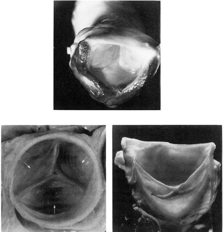
FIGURE 2. Top: The closed aortic valve viewed from below. The three coapting leaflets of the valve, the aorta above, and the ventricular myocardium along with aorto-ventricular membrane below are seen. The membrane becomes continuous with the anterior leaflet of the mitral valve. Only the load-bearing portion of the leaflet is visible. Left: The aortic view of the closed valve. Free edges of the leaflets meet at a 120° angle. Coapting surfaces, passing through the free edges, are perpendicular to the plane of the figure. The noduli are in the center and the site of leaflet attachment at their nadir is indicated by arrows. Right: A direct view of a single leaflet. The nodule of Arantius at the center of the free edge appears unusually prominent in this leaflet. The inferior margin of the lunules which is also the line of leaflet coaptation marks a separation between the coaptation surface above and the load-bearing surface below. The line of leaflet attachment to the aortic wall has a crescentic shape. (From McAlpine, W. A., Heart and Coronary Arteries, Springer-Verlag, New York, 1975. With permission.)
Excision of leaflets and sinuses can help to clarify the interrelationships of these parts. If the leaflets are cut away from the wall it can be seen that the aortic surface of each leaflet is inserted along a crescentic line that follows the proximal border of its companion sinus (Figure 3II, III). The insertion of the ventricular surface follows a different line, separated from the aortic surface by a relatively thick basal region of the leaflet. The leaflet commissures are formed by the mural regions where two leaflets insert side by side along parallel lines. The sinuses merge with each other at the commissures and continue across the sinus rim distally into the aorta (Figure 3I). Another cut, made through the wall along the curved margins of the sinuses, leaves three trigonal regions at the proximal border of the aortic root (Figure 3II, III). This scalloped border appears as a three-pointed coronet. This coronet-shaped structure has been classically, but somewhat misleadingly, called the “fibrous ring”. In fact, the fibrous elements of the root do not exist as a planar ring but form the scalloped border of a coronet. The upper borders of the sinuses also do not form a circular ring in a single plane. Instead they are scalloped (Figure 3I, II). Distally, where the leaflets come together, their line of attachment runs parallel through a certain distance. This distance will be referred to as commissural height, a term not generally found in the literature.
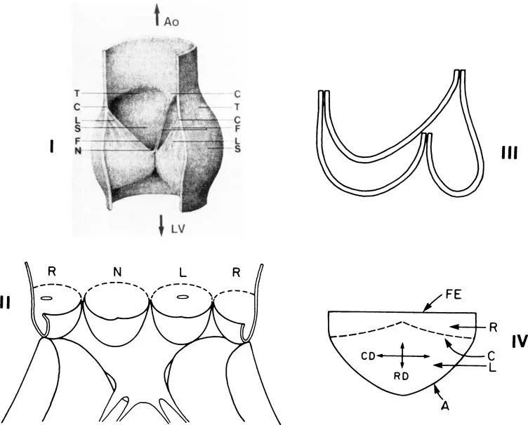
FIGURE 3. I: Aortic valve in the closed position. One leaflet and the corresponding sinus have been removed. Coronary arteries are not shown. C — commissure; F — free edge; L — lunula; N — node of Arantius; S — sinus wall, T — top of a sinus cavity. (From Sauren, A., The Mechanical Behavior of the Aortic Valve, Thesis. With permission.) II: Schematic presentation of the aortic valve opened with a longitudinal incision. L, R, and N represent left, right, and noncoronary (posterior) sinus, respectively. The dotted line shows scallop-shaped upper borders of the sinuses. The ostia of the coronary arteries are shown in R and L sinuses. (From Me Alpine, W. A., Heart and Coronary Arteries, Springer-Verlag, New York, 1975. With permission.) III: Configuration of the line of leaflet attachment to the aortic wall. IV: Drawing of a single leaflet,. FE — free edge, C — line o...
Table of contents
- Cover
- Title Page
- Copyright Page
- Dedication
- Table of Contents
- Chapter 1 Geometry of the Aortic Valve
- Chapter 2 Histology and Cytology of the Aortic Valve
- Chapter 3 Dynamics of the Aortic Valve
- Chapter 4 Fluid Dynamics of the Aortic Valve
- Chapter 5 Mechanical Stresses in the Aortic Valve
- Chapter 6 Echocardiography of the Aortic Valve
- Chapter 7 Production of Aortic Valve Sound
- Chapter 8 Diseases of the Aortic Valve
- Chapter 9 Replacement Cardiac Valves
- Appendix I
- Appendix II
- Index
Frequently asked questions
Yes, you can cancel anytime from the Subscription tab in your account settings on the Perlego website. Your subscription will stay active until the end of your current billing period. Learn how to cancel your subscription
No, books cannot be downloaded as external files, such as PDFs, for use outside of Perlego. However, you can download books within the Perlego app for offline reading on mobile or tablet. Learn how to download books offline
Perlego offers two plans: Essential and Complete
- Essential is ideal for learners and professionals who enjoy exploring a wide range of subjects. Access the Essential Library with 800,000+ trusted titles and best-sellers across business, personal growth, and the humanities. Includes unlimited reading time and Standard Read Aloud voice.
- Complete: Perfect for advanced learners and researchers needing full, unrestricted access. Unlock 1.5M+ books across hundreds of subjects, including academic and specialized titles. The Complete Plan also includes advanced features like Premium Read Aloud and Research Assistant.
We are an online textbook subscription service, where you can get access to an entire online library for less than the price of a single book per month. With over 1.5 million books across 990+ topics, we’ve got you covered! Learn about our mission
Look out for the read-aloud symbol on your next book to see if you can listen to it. The read-aloud tool reads text aloud for you, highlighting the text as it is being read. You can pause it, speed it up and slow it down. Learn more about Read Aloud
Yes! You can use the Perlego app on both iOS and Android devices to read anytime, anywhere — even offline. Perfect for commutes or when you’re on the go.
Please note we cannot support devices running on iOS 13 and Android 7 or earlier. Learn more about using the app
Please note we cannot support devices running on iOS 13 and Android 7 or earlier. Learn more about using the app
Yes, you can access The Aortic Valve by ManoJ. Thubrikar in PDF and/or ePUB format, as well as other popular books in Medicine & Cardiology. We have over 1.5 million books available in our catalogue for you to explore.