Cardiac Pacing and ICDs
eBook - ePub

Cardiac Pacing and ICDs

Kenneth A. Ellenbogen, Karoly Kaszala, Kenneth A. Ellenbogen, Karoly Kaszala

Share book
  1. English
  2. ePUB (mobile friendly)
  3. Available on iOS & Android
eBook - ePub

Cardiac Pacing and ICDs

Kenneth A. Ellenbogen, Karoly Kaszala, Kenneth A. Ellenbogen, Karoly Kaszala

Book details
Book preview
Table of contents
Citations

About This Book

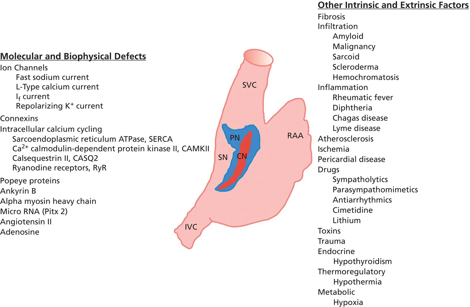
The consummate guide to cardiac pacing and defibrillator therapy in a clinical setting

Designed to provide clinicians and fellows with a complete, up-to-date breakdown of current device therapies for pacing and defibrillation, Cardiac Pacing and ICDs reflects the latest developments in the device treatment of heart rhythm abnormalities. Topics ranging from essential principals to new and innovative techniques are explored in focused chapters, illustrated with full-color images, charts, and diagrams. Addressing every aspect of permanent and temporary pacing and defibrillation therapy, this invaluable resource covers patient indications, pacing mode selection, implantation and removal techniques, troubleshooting, and much more.

The seventh edition has been expanded and revised to enable clear and practical understanding of the field as it exists today. Drawing upon real-world experience and cutting-edge research, it offers accessible, systematic guidance with a clinical focus, as well as a wealth of bitesize tips and tricks. Access to a new companion website provides insightful supplementary material, complete with downloadable images and video clips of key techniques. This essential book:

  • Provides an intuitive, easy-to-navigate guide to cardiac pacing techniques and devices
  • Explains pacing hemodynamics in practical, clinically relevant terms
  • Features simple algorithms for mode selection and device programming
  • Offers details of novel pacing systems and techniques, such as leadless pacemaker and His bundle pacing.
  • Covers pacemaker timing cycles, special features, and evaluation and management of pacing system malfunctions
  • Summarizes indications and details implantation techniques of ICDs, including transvenous and subcutaneous systems
  • Includes best practices in MRI safety, patient consultation, and remote patient follow-up

Cardiac Pacing and ICDs is an ideal resource for clinicians and fellows in cardiology and electrophysiology, those preparing for the IHRBE Examination in Devices, and any nurses, technicians, and other professionals caring for patients with implantable cardiac devices.

Frequently asked questions

How do I cancel my subscription?
Simply head over to the account section in settings and click on “Cancel Subscription” - it’s as simple as that. After you cancel, your membership will stay active for the remainder of the time you’ve paid for. Learn more here.
Can/how do I download books?
At the moment all of our mobile-responsive ePub books are available to download via the app. Most of our PDFs are also available to download and we're working on making the final remaining ones downloadable now. Learn more here.
What is the difference between the pricing plans?
Both plans give you full access to the library and all of Perlego’s features. The only differences are the price and subscription period: With the annual plan you’ll save around 30% compared to 12 months on the monthly plan.
What is Perlego?
We are an online textbook subscription service, where you can get access to an entire online library for less than the price of a single book per month. With over 1 million books across 1000+ topics, we’ve got you covered! Learn more here.
Do you support text-to-speech?
Look out for the read-aloud symbol on your next book to see if you can listen to it. The read-aloud tool reads text aloud for you, highlighting the text as it is being read. You can pause it, speed it up and slow it down. Learn more here.
Is Cardiac Pacing and ICDs an online PDF/ePUB?
Yes, you can access Cardiac Pacing and ICDs by Kenneth A. Ellenbogen, Karoly Kaszala, Kenneth A. Ellenbogen, Karoly Kaszala in PDF and/or ePUB format, as well as other popular books in Medicine & Physiology. We have over one million books available in our catalogue for you to explore.

Information

Year
2020
ISBN
9781119578284
Edition
7
Subtopic
Physiology

CHAPTER 1
Indications for permanent cardiac pacing

Roy M. John
Center for Advanced Management of Ventricular Arrhythmias, Northshore University Hospital, Manhasset, NY, USA

Introduction

Defects of cardiac impulse generation and conduction can occur at various levels in the cardiac conduction system. In general, intrinsic disease of the conduction system is often diffuse. For example, normal atrioventricular (AV) conduction cannot necessarily be assumed when a pacemaker is implanted for a disorder seemingly localized to the sinus node. Similarly, normal sinus node function cannot be assumed when a pacemaker is implanted in a patient with AV block. Conduction disorders that lead to important bradycardia or asystole may result from reversible or irreversible causes. Recognition of reversible causes is critical to avoid unnecessary commitment to long‐term pacemaker therapy. This chapter reviews the common disorders that warrant cardiac pacing and lists the recommended indications set out by published guidelines.

Anatomy and physiology of the conduction system

For a complete understanding of rhythm generation and intracardiac conduction, and of their pathology, a brief review of the anatomy and physiology of the specialized conduction system is warranted.

Sinus node

The sinus node or sinoatrial (SA) node is a crescent‐shaped subepicardial structure located at the junction of the right atrium and superior vena cava along the terminal crest. It measures 10–20 mm (with larger extension in some studies) and has abundant autonomic innervation and blood supply, with the sinus node artery commonly coursing through the body of the node. Endocardially, the crista terminalis overlies the nodal tissue, although the inferior aspect of the node has a more subendocardial course. Histologically, the sinus node comprises specialized nodal cells (P cells) packed within a dense matrix of connective tissue. At the periphery, these nodal cells intermingle with transitional cells and the atrial working myocardium, with radiations extending toward the superior vena cava, the crista terminalis, and the intercaval regions [1,2]. The absence of a distinct border and the presence of distal fragmentation explain the lack of a single breakthrough of the sinus node excitatory wavefront. The radiations of the node, although histologically distinct, are not insulated from the atrial myocardium. Hence, a clear anatomical SA junction is absent. The sinus node is protected from the hyperpolarizing effect of the surrounding atria, probably by its unique structure wherein electrical coupling between cells and expression of ion channels vary from the center of the node to the periphery. The pacemaker cells at the center of the node are more loosely coupled, while those at the periphery are more tightly coupled with higher density If (funny current, a mixed sodium and potassium current carried by the HCN channels) and INa currents [2].
The SA node has the highest rate of spontaneous depolarization (automaticity) in the specialized conduction system and is responsible for the generation of the cardiac impulse under normal circumstances, although normal human pacemaker activity may be widely distributed in the atrium. The unique location of the sinus node astride the large SA nodal artery provides an ideal milieu for continuous monitoring and instantaneous adjustment of heart rate to meet the body’s changing metabolic needs.
Impulse generation in the sinus node remains incompletely understood. Sinus nodal cells have a low resting membrane potential of −50 to −60 mV. Spontaneous diastolic (phase 4) depolarizations are probably triggered by several currents, including If. The predominant inward current in the center of the node is ICaL that generates a “slow” action potential. The action potentials spread peripherally into the musculature of the terminal crest. In the periphery of the node, INa is operative and necessary for providing sufficient inward current to depolarize the larger mass of atrial tissue. Defects of a number of molecular and biophysical factors that govern the ionic channels of the sinus node can lead to sinus node dysfunction (Figure 1.1).
Differential sensitivity to adrenergic and vagal inputs exists along the nodal pacemaker cells, such that superior sites tend to dominate during adrenergic drive while the inferior sites predominate during vagal stimulation [3]. Interventions including premature stimulation, autonomic stimulation, and drugs have been shown to induce pacemaker shifts (due to multicentric origins) with variable exit locations [4].
Diagram of the right atrium of the heart displaying the central node, sinus node, etc. The molecular and biophysical defects are listed at the left, while the other intrinsic and extrinsic factors are listed at the right.
Figure 1.1 Summary of factors contributing to sinus node (SN) dysfunction. The central node (CN) shown in red is surrounded by the peripheral nodal (PN) structure in blue. RAA, right atrial appendage; SVC, superior vena cava; IVC, inferior vena cava.
Source: modified from Monfredi O, Boyett MR. Sinus sinus syndrome and atrial fibrillation in older persons: a view from the sinoatrial nodal myocyte. J Mol Cell Cardiol 2015;83:88–100. Reproduced with permission of Elsevier.

Atrioventricular node

The compact AV node is a subendocardial structure situated within the triangle of Koch and measuring 5–7 mm in length and 2–5 mm in width [5,6]. On the atrial side, the node is an integral part of the atrial musculature, in contrast to the AV bundle which is insulated within the central fibrous body and merges with the His bundle. Based on action potential morphology in rabbit hearts, atrial (A), nodal (N), and His (H) cells have been defined. Intermediate cell types such as AN and NH define areas toward the atrial and His bundle ends of the compact node, respectively. Histologically, the mid nodal part has densely packed cells in a basket‐like structure interposed between the His bundle and the loose atrial approaches to the node. The AN cells are composed primarily of transitional cells. Distinct electrical and morphological specialization is seen only in the progressively distal His fibers. Rightward and leftward posterior extensions of the AV node were described by Inoue and Becker [7]. These extensions have clinical implications for defining reentrant circuits that act as a substrate of AV nodal reentrant tachycardia.
The AV node has ext...

Table of contents