- 259 pages
- English
- ePUB (mobile friendly)
- Available on iOS & Android
eBook - ePub
Dirofilariasis
About this book
Heartworm disease is probably the most well-known disease of dogs, and its epidemiology is being recorded in those countries in which the disease is routinely treated in the domestic dog. It is hoped that this text will be of benefit to us all and that our understanding of this disease will continue to grow and so enable us to resolve some of the exsiting problems associated with the treatment of this disease. To this end, the book aims to project some hypotheses in different areas that, while unproven, seem to be the most probable, based on our current knowledge.
Trusted by 375,005 students
Access to over 1.5 million titles for a fair monthly price.
Study more efficiently using our study tools.
Information
Chapter 1
Dirofilaria Sp.: Taxonomy and Distribution
James B. Lok
TABLE OF CONTENTS
- I. Systematics
- II. Morphology
- A. Adults
- 1. Gross Morphology
- 2. Morphology Apparent in Tissue Sections
- B. Microfilariae
- III. Geographic Distribution
- A. North America
- B. South America
- C. Europe
- D. Africa
- E. Asia and the Pacific
- F. Australia
- G. The Changing World Distribution of Dirofilaria immitis
- IV. Filarioids Other than Dirofilaria immitis in Dogs and Cats
- A. Dipetalonema reconditum
- B. The “Irish” Dipetalonema
- C. Dirofilaria striata
- D. The “Florida” Dirofilaria
- V. Vectors of Dirofilaria immitis
- Acknowledgments
- References
I. Systematics
Dirofilaria immitis, as the agent of canine cardiovascular dirofilariasis, is a parasite of paramount veterinary importance. It is increasingly recognized as the cause of zoonotic infections, most frequently as the agent of human pulmonary dirofilariasis.1 The parasite, now known to occur throughout the world in many species of host, was first described in 1850 in Philadelphia, by physician J. Leidy who assigned the name Filaria canis cordis.2 The worm was renamed Filaria immitis by Leidy in 1856.3 and the genus Dirofilaria was erected in 1911 by Railliet and Henry4 with Filaria immitis as its type species.2 Synonyms in addition to those listed above include Filaria spirocauda, F. haematica, F. sanguinis, F. hebetata, Dirofilaria nasuae, D. pongoi, D. fausti, D. louisianensis, D. indica,5 and D. magalhaesi.1 The taxonomy of the genus Dirofilaria has been reviewed by Anderson.6
Like other members of the superfamily Filarioidea the genus Dirofilaria consists of elongate, thin nematodes which live outside the alimentary tract and which are viviparous giving birth to motile first-stage larvae called microfilariae.1 As is the case with all filariae, members of this genus require an arthropod intermediate host, generally a mosquito or black fly, to complete their life cycles.5–7 The species generally recognized as being of primary veterinary importance belong to two subgenera. The subgenus Dirofilaria contains those worms such as D. immitis which have smooth cuticles and which, as adults, are usually found in the cardiovascular system. Members of the subgenus Nochtiella such as D. ursi, D. repens, D. striata, and D. tenuis generally dwell in subcutaneous tissues of the definitive host and have prominent longitudinal ridges and fine striations on their cuticles.1 These differential subgeneric characters are discussed more fully below.
II. Morphology
A. Adults
1. Gross Morphology
The genus Dirofdaria consists of elongate, thin filarioid nematodes, whitish in coloration, with bluntly rounded anterior extremities, rudimentary buccal capsules without lips, small cephalic papillae and an abbreviated esophagus indistinctly differentiated into muscular and glandular regions (Figure 1A, B). The caudal extremities of female worms are also bluntly rounded (Figure 1E), and the vulvar openings are located just posterior to the junction of the esophagus and intestine. The caudal ends of males are coiled spirally and are more conical in shape then those of females. In all species of Dirofilaria spicules are unequal in length (Figure 1C). The gubernaculum is absent.5,6
Dirofdaria (Dirofilaria) immitis is the longest of the Dirofilaria spp. of medical and veterinary importance. Females measure 250 to 310 mm in length and 1.0 to 1.3 mm in width. They have an obtuse caudal end with the vulvar opening some 2.7 mm from the anterior end. Males of D. immitis are 120 to 200 mm long and 0.7 to 0.9 mm wide. The spirally coiled caudal end bears five pairs of preanal and six pairs of postanal papillae (Figure 1C). The left spicule is 300 to 375 µm long, and the right 175 to 299 µm. In both sexes the stoma is surrounded externally by six inconspicuous papillae (Figure 1B).5
Other species of Dirofdaria which occur in dogs and cats and which may occasionally affect man are readily distinguished from D. immitis on the basis of size. Females of Dirofilaria repens, a parasite of the subcutaneous tissues of dogs and cats throughout Africa, Southern Europe, India, and Southeast Asia, measure 100 to 170 mm in length and 0.46 to 0.65 mm in width. Males are 50 to 70 mm long and 0.37 to 0.45 mm wide with two to six preanal papillae to the right of the anus and four to five to the left. The spicules are highly dissimilar, the left being 460 to 590 µm long and the right being 180 to 210 µm long. Microfilariae of D. repens occur in the subcutaneous lymph spaces and in the peripheral blood. On the basis of size alone it would appear difficult to distinguish the microfilariae of D. repens from those of D. immitis. The range of lengths, 268 to 360 µm, reported for these parasites overlaps that reported for D. immitis microfilariae. The relative positions of somatic structures such as the nerve ring, the excretory cell, the G1 cell, and the anal space (Figure 1F) along the length of the microfilaria provide some means of diagnosis.
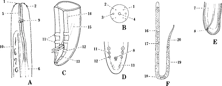
FIGURE 1. Gross morphology of D. immitis. (A) Anterior end of adult female showing (1) inner circle of cephalic papillae, (2) outer circle of cephalic papillae, (5) muscular esophagus, (6) glandular esophagus. (9) nerve ring and (10) uterus. (B) En face view of anterior end of adult worm showing (1) inner and (2) outer circles of cephalic papillae, (3) amphids, and (4) stoma. (C) Caudal end of adult male showing (11) preanal papillae, (12) adanal papillae, (13) postanal papillae. (14) long spicule. (15) and short spicule. (D) Ventral view of caudal end of male showing (11) preanal. (12) adanal, and (13) postanal papillae and (8) anus. (E) Caudal view of adult female showing (7) intestine and (8) anus. (F) Microfilaria from peripheral blood showing (16) nerve ring, (17) excretory cell. (18) G1 cell, (19) anal space, (20) and tail cells. (From Olsen, O. W., Animal Parasites, Their Life Cycles and Ecology, 3rd ed., University Park Press, Baltimore, 1974, 491. With permission.)
2 Morphology Apparent in Tissue Sections
Human pulmonary infections with Dirofilaria immitis and subcutaneous infections with D. tenuis, D. repens, and D. ursi are being reported with increasing frequency8 (see also Boreham, chapter 13). Moreover, D. immitis has been reported from numerous ectopic locations in both dogs and alternative, definitive, and aberrant hosts.9,10 For these reasons the clinician may be required to recognize the characteristics of the parasite in tissue cross sections. Gutierrez8 describes the characteristics of adult Dirofilaria spp. as seen in histological sections. Adults of the genus Dirofilaria have relatively thick multilaminate cuticles with individual layers most apparent in proximity to the lateral chords (Figures 2 and 3). The external surfaces of the cuticles of D. ursi, D. tenuis, and D. repens bear longitudinal ridges which are prominent in cross sections. The cuticle of D. immitis, on the other hand, is relatively smooth with longitudinal ridges occurring only in the caudal area on the ventral aspect. Two of the cuticular layers are fibrous in structure with fibers of one layer running perpendicular to those of the other and the whole array running oblique to the axis of the body. These cuticular fibers appear as a criss-cross network when viewed in tangential sections of adult worms. The lateral cords are prominent in cross sections with well-developed musculature and numerous cells (Figure 3).
Identification of Dirofilaria spp. in cross section is done primarily on the basis of structure and arrangement of the longitudinal cuticular ridges. Longitudinal ridges of adult D. repens are conspicuous and sharp, separated by a space three to four times the width of the ridge itself. Dirofilaria tenuis has a distinctive pattern of low, rounded ridges in a branching network with spaces appearing narrower than the ridges themselves. As mentioned above, the cuticle of D. immitis is smooth with...
Table of contents
- Cover
- Title Page
- Copyright Page
- Table of Contents
- Chapter 1 Dirofilaria Sp.: Taxonomy and Distribution
- Chapter 2 Biology of Dirofilaria Immitis
- Chapter 3 The Biochemistry of Dirofilaria Immitis
- Chapter 4 Clinical Signs and Diagnosis of Canine Dirofilariasis
- Chapter 5 Radiology of Heartworm Disease
- Chapter 6 Pathology and Pathogenesis of Dirofilariasis
- Chapter 7 Therapy in Relation to Aspects of the Pathophysiology of Dirofilariasis
- Chapter 8 Chemotherapy and Chemoprophylaxis
- Chapter 9 Adverse Reactions to Diethylcarbamazine in the Treatment of Dirofilariasis
- Chapter 10 Immunology of Canine Dirofilariasis
- Chapter 11 Caval Syndrome
- Chapter 12 Feline Heartworm Disease
- Chapter 13 Dirofilariasis in Man
- Appendix
- Index
Frequently asked questions
Yes, you can cancel anytime from the Subscription tab in your account settings on the Perlego website. Your subscription will stay active until the end of your current billing period. Learn how to cancel your subscription
No, books cannot be downloaded as external files, such as PDFs, for use outside of Perlego. However, you can download books within the Perlego app for offline reading on mobile or tablet. Learn how to download books offline
Perlego offers two plans: Essential and Complete
- Essential is ideal for learners and professionals who enjoy exploring a wide range of subjects. Access the Essential Library with 800,000+ trusted titles and best-sellers across business, personal growth, and the humanities. Includes unlimited reading time and Standard Read Aloud voice.
- Complete: Perfect for advanced learners and researchers needing full, unrestricted access. Unlock 1.5M+ books across hundreds of subjects, including academic and specialized titles. The Complete Plan also includes advanced features like Premium Read Aloud and Research Assistant.
We are an online textbook subscription service, where you can get access to an entire online library for less than the price of a single book per month. With over 1.5 million books across 990+ topics, we’ve got you covered! Learn about our mission
Look out for the read-aloud symbol on your next book to see if you can listen to it. The read-aloud tool reads text aloud for you, highlighting the text as it is being read. You can pause it, speed it up and slow it down. Learn more about Read Aloud
Yes! You can use the Perlego app on both iOS and Android devices to read anytime, anywhere — even offline. Perfect for commutes or when you’re on the go.
Please note we cannot support devices running on iOS 13 and Android 7 or earlier. Learn more about using the app
Please note we cannot support devices running on iOS 13 and Android 7 or earlier. Learn more about using the app
Yes, you can access Dirofilariasis by P. F. L. Boreham in PDF and/or ePUB format, as well as other popular books in Medicine & Veterinary Medicine. We have over 1.5 million books available in our catalogue for you to explore.