- 520 pages
- English
- ePUB (mobile friendly)
- Available on iOS & Android
eBook - ePub
Immunology of Insects and Other Arthropods
About this book
In insect and other arthropod immune systems, discrimination between self and nonself tissues is accomplished through the combined actions of two immunocytes and several humoral factors. Immunology of Insects and Other Arthropods presents a comprehensive look at this and other important topics in arthropod immunology. Issues discussed include insect immunocytes and other hemocytes, including computer image analysis of immunocyte serial sections; the two basic cellular immune reactions (phagocytosis and encapsulation), including the molecular basis and roles of gap junctions in encapsulation; how encapsulation is affected by polydnavirus and encapsulation-promoting factors; why insect cells are immune to HIV; humoral factors; and antibacterial factors in Lepidoptera, Diptera, and other insect orders.
Other topics include hemolymph proteins interacting with mammalian complement cascade; adaptive humoral response in the American cockroach; antigenic stimulation of hemaglutinin production in insects; and the applications of the Limulus Amebocyte Lysate (LAL) in detecting endotoxins in pharmaceuticals, medical devices, clinical diagnosis, and hygienic control. This book represents an important reference source for hematologists, pathologists, immunologists, AIDS researchers, comparative immunologists, and pharmaceutical companies.
Trusted by 375,005 students
Access to over 1.5 million titles for a fair monthly price.
Study more efficiently using our study tools.
Information
Immunocytes and Their Immune Reactions
Chapter 1
Cellular and Humoral Immunity in the Horseshoe Crab, Limulus Polyphemus
Peter B. Armstrong
TABLE OF CONTENTS
- I. Introduction
- II. Pathogenic Challenge in Limulus
- III. Characterization of the Hemocytes of Limulus
- IV. The Activation Reaction
- V. Phagocytosis
- VI. Other Cell-Mediated Defense Reactions
- VII. Humoral Defense Systems
- VIII. Summary
- Acknowledgments
- References
I. Introduction
Individual metazoans are threatened by a nearly continual challenge by microbial and metazoan pathogens. The life cycle time for most microbial pathogens is significantly shorter than that of the metazoan host, making it essentially impossible for the host population to outgrow populations of the pathogens. Instead, metazoans have evolved a varied array of defense strategies that limit their susceptibility to attack by potential pathogens. These include the presence of surface tissues that restrict penetration of pathogens into the interior, and a variety of specialized cells and humoral factors that fight pathogens which have succeeded in gaining access into the body. In higher animals, a majority of the internal defense systems involve cells and soluble factors of the blood. This chapter will review the defense systems of the arthropod, Limulus, with particular emphasis on the involvement of the blood cells and blood-borne soluble factors in defense.
II. Pathogenic Challenge in Limulus
Several pathogens of Limulus have been identified. Strains of the Gram-negative marine bacterium Vibrio have been isolated from moribund animals.1 The animal is also subject to cutaneous infection by a variety of organisms, including filamentous and unicellular cyanobacteria2 that eventually penetrate the cuticle and provide a pathway for invasion by other bacteria. The triclad turbellarid worm, Bdelloura candida, which is often abundant in the gills, causes lesions in the cuticle that are portals for entry of microbial pathogens into the body.3, 4 and 5 Although Limulus lives in a septic environment and is subject to frequent breach of the surface integument, the hemolymph of a majority of recently collected animals is sterile.6 Presumably, invading pathogens are effectively controlled by the internal defenses. These defense systems will be the subject of this review.
III. Characterization of the Hemocytes of Limulus
Limulus is one of the largest, easily obtainable arthropods. The blood occupies approximately one-third of its body mass. It is relatively easy to obtain 100 or more milliliters of blood from an average-sized adult.7 Limulus is, thus, an ideal subject for hematologic studies requiring large volumes of material. Based on cell morphology, there appears to be only one blood cell type in the systemic circulation of the adult intermolt animal — the granulocyte or amebocyte.8, 9, 10, 11 and 12 This cell is an oval, plate-shaped structure, 15 to 20 μm in its longest dimension (Figure 1). The cytoplasm is packed with large, highly refractile exocytotic granules. The discoid shape is stabilized by a circumferential bundle of microtubules.13 The amebocyte is stimulated by extravasation to become motile14, 15, 16, 17 and 18 and adhesive15,19 and 20 and to undergo degranulation.10,21, 22, 23 and 24 The alterations in morphology attendant with these responses result in a pleiomorphic population of cells that are very different in appearance from the unstimulated circulating cells, but nevertheless are derived from them16, 17 and 18,25 (Figure 2). This variation in cell morphology led some investigators to postulate the existence of multiple cell types in the blood.26,31 However, careful study of both living and quickly fixed samples has convinced us that the peripheral blood of the adult contains but one cell type, with the more overt morphological heterogeneity ascribable to alterations resultant from cellular activation. Some degree of heterogeneity, such as a variable content of autofluorescent cytoplasmic granules11 and electron-dense inclusions,12 is real, but of unknown significance. Limulus does have a second blood cell, the cyanocyte,32,33 but in the adult this cell is restricted to the vascular spaces of the ocular ganglion, and is apparently absent from the general circulation of the adult, intermolt animal.11,34 The cyanoblast does appear, however, to be systemically distributed in the early larval stages.35
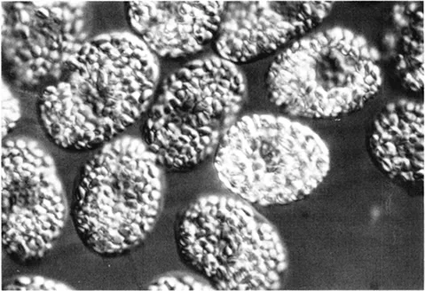
Figure 1 Limulus blood cells in the oval configuration characteristic of unextravasated blood. The cells are oval, nonadhesive, immobile, and fully granular. Cell morphology and behavior change dramatically upon the transformation that follows removal of the cells from the animal. (× 1243) (From Armstrong, P. B., Exp. Cell Res., 107, 127, 1977. With permission.)
IV. The Activation Reaction
As indicated above, the circulating amebocyte is an ovoid, granular, nonadhesive, and nonmotile cell. At the morphological level, the cell activation that follows extravasation establishes an alternative phenotype, in which the cell becomes pleiomorphic, adhesive, and motile, and from which the contents of the cytoplasmic granules are released into the external milieu by exocytosis. Limulus lacks the explosive blood cells of crustaceans, which respond to extravasation by cytolysis (reviewed in Hose et al.36). Instead, the extravasated Limulus blood cells survive,16,17,37,38 but lose the cytoplasmic granules by exocytosis. The amebocyte is responsive to bacterial products, such as lipopolysaccharide (LPS, endotoxin — LPS is the principal toxin of Gram-negative bacteria).1,21, 22 and 23,39, 40, 41 and 42 The cells also activate, albeit more slowly, if prepared under LPS-free conditions,17,23,25 indicating the existence of LPS-inde-pendent activation pathways.
Relatively little is known of the pathways that couple initial external stimulation with the family of intracellular responses that characterize the activated state. Activation is significantly retarded in the absence of extracellular divalent cations,23,43 which suggests that movement of divalent cations into the cell may be involved. In this regard, Mg2+ appears to be more effective than Ca2+ in stimulating isolated cells to degranulate.44 Presumably second messengers, such as cyclic nucleotides and/or the metabolic products of phospho-inositide breakdown, are involved, possibly acting through membrane receptors for LPS.45,46 Cyclic-AMP appears to be involved, although, whether in the potentiation or in the inhibition of exocytosis is controversial. Agents that elevate c-AMP levels have been variously reported to retard23 or potentiate47 activation. The acquisition of locomotory capabilities and adhesiveness, and the extrusion of the cytoplasmic granules by exocytosis all imply that activation is accompanied by reorganizations of the cytoskeleton and alteration of the composition of the cell surface. Bundles of aligned actin filaments appear in the filopodial extensions of motile cells.11,48 Surprisingly, a circumferential bundle of microtubules, which apparently stabilizes the discoidal shape of the unactivated cell, remains intact, at least through the early stages of activation, although it may be displaced from its original sub-plasmalemmal location.48 It has been proposed that the aggregation of the amebocytes is mediated by an interaction between a cell surface hemagglutinin and coagulogen bound to the cell surface,43,49,50 but this proposal has not been rigorously proved. (Coagulogen is the structural protein of the extracellular blood clot in Limulus.)
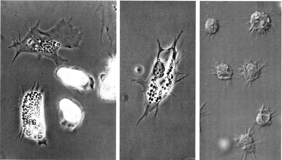
Figure 2 Extravasated blood cells adherent to a microscopic coverglass showing the range of phenotypes adopted by cells under these conditions. In the left panel, the two cells in the lower right are compact and highly motile, with hyaline pseudopods at the anterior ends. The cell in the lower left is partially flattened and fully granular. Such cells are motile and may revert to the highly motile phenotype. The cell in the upper center is highly flattened on the substratus and has suffered exocytosis of all but two of its complement of cytoplasmic granules. (From Armstrong, P. B., Exp. Cell Res., 107, 127, 1977. With permission.) In the middle panel, the flattened cell is in the process of degranulation, with the large clear region in the center being the site of recent exocytosis. (From Armstrong, P. B., Blood Cells of Marine Invertebrates: Experimental Systems in Cell Biology, W. D. Cohen, Ed., Alan R. Liss, p. 77. With permission.) The cells of the right panel have been stimulated to degranulate by exposure to bacterial lipopolysaccharide. From Armstrong, P. B., Blood Cells of Marine Invertebrates: Experimental Systems in Cell Biology, W. D. Cohen, Ed., Alan R. Liss. p. 77. With permission.) (× 1046)
The functional significance of activation is more clear. One of the major functions of the granular amebocyte is the regulation of hemostasis. Two of the principal requirements of any hemostatic system are that it be inactive in the intact animal and that it be responsive to bleeding by a rapid activation that is spatially restricted to the wound site. The activation response of the granular amebocyte clearly fits these requirements. The acquisition of cellular adhesiveness allows the formerly nonadhesive amebocytes to attach to the borders of the wound and to one another to form a cellular hemostatic plug that arrests bleeding.14,19,26,51,52 In addition to the cellular clot, the hemostatic response includes the formation of an extracellular clot,42,53 in which the amebocytes play a dominant role. The extracellular clot is composed of fibers of the protein coagulin.54,64 In the intact animal, the soluble precursor of this protein, coagulogen, is absent from the plasma, but is instead sequestered in the cytoplasmic granules of the amebocytes.65,67 Granule exocytosis releases coagulogen into the external milieu. Coagulogen is converted to coagulin by a proteolytic cleavage administered by a particular serine protease, known as clotting e...
Table of contents
- Cover
- Title Page
- Copyright Page
- Contents
- Series Preface
- Preface
- The Editor
- Contributors
- Section I. Immunocytes and Their Immune Reactions
- Section II. Coagulation
- Section III. Humoral Factors
- Section IV. Biomedical Applications and Techniques
- Taxonomic Index
- Subject Index
Frequently asked questions
Yes, you can cancel anytime from the Subscription tab in your account settings on the Perlego website. Your subscription will stay active until the end of your current billing period. Learn how to cancel your subscription
No, books cannot be downloaded as external files, such as PDFs, for use outside of Perlego. However, you can download books within the Perlego app for offline reading on mobile or tablet. Learn how to download books offline
Perlego offers two plans: Essential and Complete
- Essential is ideal for learners and professionals who enjoy exploring a wide range of subjects. Access the Essential Library with 800,000+ trusted titles and best-sellers across business, personal growth, and the humanities. Includes unlimited reading time and Standard Read Aloud voice.
- Complete: Perfect for advanced learners and researchers needing full, unrestricted access. Unlock 1.5M+ books across hundreds of subjects, including academic and specialized titles. The Complete Plan also includes advanced features like Premium Read Aloud and Research Assistant.
We are an online textbook subscription service, where you can get access to an entire online library for less than the price of a single book per month. With over 1.5 million books across 990+ topics, we’ve got you covered! Learn about our mission
Look out for the read-aloud symbol on your next book to see if you can listen to it. The read-aloud tool reads text aloud for you, highlighting the text as it is being read. You can pause it, speed it up and slow it down. Learn more about Read Aloud
Yes! You can use the Perlego app on both iOS and Android devices to read anytime, anywhere — even offline. Perfect for commutes or when you’re on the go.
Please note we cannot support devices running on iOS 13 and Android 7 or earlier. Learn more about using the app
Please note we cannot support devices running on iOS 13 and Android 7 or earlier. Learn more about using the app
Yes, you can access Immunology of Insects and Other Arthropods by Ayodhya P. Gupta in PDF and/or ePUB format, as well as other popular books in Biological Sciences & Biology. We have over 1.5 million books available in our catalogue for you to explore.