Machine Vision systems combine image processing with industrial automation. One of the primary areas of application of Machine Vision in the Industry is in the area of Quality Control. Machine vision provides fast, economic and reliable inspection that improves quality as well as business productivity. Building machine vision applications is a challenging task as each application is unique, with its own requirements and desired outcome.
A Guide to Machine Vision in Quality Control follows a practitioner's approach to learning machine vision. The book provides guidance on how to build machine vision systems for quality inspections. Practical applications from the Industry have been discussed to provide a good understanding of usage of machine vision for quality control. Real-world case studies have been used to explain the process of building machine vision solutions.
The book offers comprehensive coverage of the essential topics, that includes:
Introduction to Machine Vision
Fundamentals of Digital Images
Discussion of various machine vision system components
Digital image processing related to quality control
Overview of automation
The book can be used by students and academics, as well as by industry professionals, to understand the fundamentals of machine vision. Updates to the on-going technological innovations have been provided with a discussion on emerging trends in machine vision and smart factories of the future.
Sheila Anand is a PhD graduate and Professor at Rajalakshmi Engineering College, Chennai, India. She has over three decades of experience in teaching, consultancy and research. She has worked in the software industry and has extensive experience in development of software applications and in systems audit of financial, manufacturing and trading organizations. She guides Ph.D. aspirants and many of her research scholars have since been awarded their doctoral degree. She has published many papers in national and international journals and is a reviewer for several journals of repute.
L Priya is a PhD graduate working as Associate Professor and Head, Department of Information Technology at Rajalakshmi Engineering College, Chennai, India. She has nearly two decades of teaching experience and good exposure to consultancy and research. She has delivered many invited talks, presented papers and won several paper awards in International Conferences. She has published several papers in International journals and is a reviewer for SCI indexed journals. Her areas of interest include Machine Vision, Wireless Communication and Machine Learning.
Trusted by 375,005 students
Access to over 1.5 million titles for a fair monthly price.
Computer vision (CV) has evolved into a major discipline that involves both cameras and computing technologies. However, like all major technologies, the beginning was albeit a small one. In the summer of 1966, Seymour Papert and his colleague Marvin Minsky of the Artificial Intelligence (AI) Laboratory at MIT were assigning the Summer Vision Project to undergraduates. The aim of the project was to build a system that could analyze a scene and identify objects in it. Marvin Minsky was said to have famously instructed a graduate student to “connect a camera to a computer and have it describe what it sees.” However, as it turned out, it was easier said than done. Both Papert and Minsky went on to become pioneers in the fields of artificial intelligence and computer vision and have paved the way for considerable research in these respective fields.
Computer vision is the field of science/technology that deals with providing computers with vision; simply stated, how computers can, like humans, see and understand real-world objects. For instance, when humans see a flower, we can immediately perceive that it is a flower. How do we identify it? From the shape, color, smell, or all of it? So, would anything that has shape, color, and smell be a flower? That would not be true as there are other objects, sweets for example, that have shape, color, and smell. In other words, how is the human brain able to process the image of the flower that it sees and understand it as a flower? Computer vision, therefore, is not merely duplicating human vision but also the ability to process and interpret the image.
Computer vision systems use cameras to capture images in the real world. The captured image is then analyzed to understand the image so that appropriate decisions or suitable actions can be initiated. For example, the image may be captured and analyzed to determine the distance between the camera and a building for route mapping. In autonomous vehicle management, computer vision could be used to track the path of a vehicle to determine if it is being driven in the middle of its lane. Another typical example is determining the number of people present in a stadium for crowd management. In a totally different arena, a plate of food could be analyzed to find out the type and quantity of food present as well as its nutritive value. The possibilities are enormous and computer vision can virtually be applied to any field to initiate and perform various actions.
But our focus in this book is machine vision (MV). So, what then is machine vision? Computer vision covers the core technology of image analysis that can be used for any application. Machine vision, on the other hand, usually refers to a process of combining image analysis with industrial automation. For example, machine vision could be applied for automated inspection or robot guidance in industrial applications. Likewise, in process control, the parts, subassemblies, assemblies, or end products can be automatically inspected to find manufacturing defects. It is easy to see that there is considerable overlay between the fields of computer vision and machine vision.
1.1 The Human Eye
Computer vision tries to mimic the human vision system. So, we first need to understand how the human eye captures and processes images. Vision is one of most advanced of the human senses and the eyes are responsible for vision and nonverbal communication in the human vision system. The human eye uses a single lens to focus images onto a light-sensitive membrane called the retina. The obtained image is then sent to the brain for processing. The brain analyzes the images and initiates suitable action. Referring to our earlier example, once the brain identifies an image as a flower, the outcome could be simply to appreciate the beauty of the flower or action may be initiated to smell or pluck the flower.
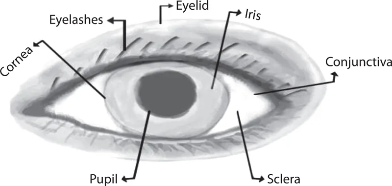
We will now look at the anatomy of the human eye to learn about the different parts of the eye and understand how they work together to provide humans with vision. As the eyes are one of the most vital sense organs in the human body, they must be well protected from dust, dirt, and injuries that will affect human vision. The eye is enclosed in a socket that protects the eye from mechanical and other injuries. The socket is made up of parts of several bones of the skull to form a four-sided pyramid with the apex pointing to the back into the head. The eyeball and its functional muscles are surrounded by a layer of orbital fat that acts like a cushion and permits the eyeball to rotate smoothly around a virtual pivotal point. The outer structure of the human eye is shown in Figure 1.1.
The eye has several more layers of protection that safeguard the human vision system. Eyelashes protect the eye and prevent dust and other small particles like sand from entering the eye. The eyelashes are also highly sensitive and provide a warning when an object comes too close to the eye. Another notable feature is that the upper set of eyelashes curves upward, while the lower set of eyelashes curves downward to prevent the two sets of eyelashes from interlacing. In terms of vision, they regulate the eye from sunshine to a certain extent and provide the first layer of protection.
FIGURE 1.1 Outer structure of the human eye.
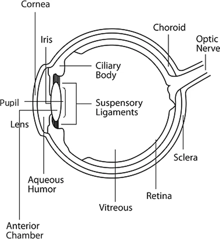
The next layer that protects the eye are the eyelids, which are movable folds of tissues. There are several muscles that help the eye to blink and help to keep our eyes open. When we blink our eye, tears are secreted, and the eyelids help to spread the tears evenly over the surface of the eye to keep it moist and lubricated. A simplified inner structure of the eye is shown in Figure 1.2.
The sclera is the thick white part of the eye also known as the “white of the eye.” It covers the surface of the eyeball and extends all the way to the back of the eye to the optic nerve. It is opaque and fibrous and provides strength and protection to the eye. Its outer surface is covered by a thin vascular covering called the episclera. The collagen bundles in sclera are of varying sizes and are irregularly arranged; hence, the sclera is not transparent like cornea. The sclera provides a sturdy attachment for the extraocular muscles that control the movement of the eyes.
FIGURE 1.2 Inner structure of the human eye.
The sclera surrounds the cornea, which is the clear dome-shaped surface that lies in the front of the eye and covers the iris, pupil, and anterior chamber. The cornea admits and helps to focus light waves as they enter the eye. The cornea is avascular, which means that it gets no blood supply. It absorbs oxygen from the air and receives its nourishment from tears and the aqueous humor, which is a fluid in the front part of the eye that lies behind the cornea. The tissues of the cornea are arranged in three basic layers, with two thinner layers, or membranes, between them. Each of these five layers has an important function. The epithelium is the cornea’s outermost layer. It acts as a barrier to prevent dust, water, and other foreign particles from entering the eye. The epithelium is filled with thousands of tiny nerve endings, which is why your eye may hurt when it is rubbed or scratched. The part of the epithelium that epithelial cells anchor and organize themselves to is called the basement membrane. The next layer behind the basement membrane of the epithelium is a transparent film of tissue called Bowman’s layer, composed of protein fibers called collagen. If injured, Bowman’s layer can form a scar as it heals. If these scars are large and centrally located, they may cause vision loss. Behind Bowman’s layer is the stroma, which is the thickest layer of the cornea. It is composed primarily of water and collagen. Collagen gives the cornea its strength, elasticity, and form. The unique shape, arrangement, and spacing of collagen proteins are essential in producing the cornea’s light-conducting transparency. Behind the stroma is Descemet’s membrane, a thin but strong film of tissue that serves as a protective barrier against infection and injuries. Descemet’s membrane is composed of collagen fibers that are different from those of the stroma and are made by cells in the endothelial layer of the cornea. The endothelium is the thin, innermost layer of the cornea. Endothelial cells are important in keeping the cornea clear. Normally, fluid leaks slowly from inside the eye into the stroma. The endothelium’s primary task is to pump this excess fluid out of the stroma. Without this pumping action, the stroma would swell with water and become thick and opaque. In a healthy eye, a perfect balance is maintained between the fluid moving into the cornea and the fluid pumping out of the cornea. Unlike the cells in Descemet’s membrane, endothelial cells that have been destroyed by disease or trauma are not repaired or replaced by the body. The cornea was one of the first organs to be successfully transplanted because it lacks blood vessels.
The conjunctiva is a thin, translucent membrane that lines the inside of the eyelids and covers the sclera. The conjunctiva folds over itself to allow unrestricted eyeball movement. The conjunctiva has its own nerve and blood supply. The main function of the conjunctiva is to keep the eye lubricated by producing mucus and some level of tears. It protects the eye by preventing microbes and other foreign particles from entering the eye. The limbus forms the border between the transparent cornea and opaque sclera.
The pupil is the black circle in the center of the eye, and its primary function is to monitor the amount of light that comes into the eye. When there is a lot of light, the pupil contracts to keep the light from overwhelming the eye. When there is very little light, the pupil expands so it can soak up as much light as possible. The iris is the colored part of the eye. The iris functions to adjust the size of the pupil. It has muscles that contract or expand depending on the amount of light the pupil needs to process images.
The lens lies behind the pupil and is responsible for allowing the eyes to focus on small details like words in a book. The lens is in a constant state of adjustment as it becomes thinner or thicker to accommodate the detailed input it receives. With age, the lens loses a lot of its elasticity, which often results in cataracts and other eye conditions because the lens cannot adjust as well to its surroundings as it used to.
The space between the cornea and the iris is known as the anterior chamber. The posterior chamber is the space between the iris and lens. These chambers are filled with a transparent, watery fluid known as aqueous humour, which is similar to plasma but contains low protein concentrations and is secreted from the ciliary epithelium, which is a structure that supports the lens.
The retina is the light-sensitive membrane that lines the inner surface of the back of the eyeball. Images are formed on the retina and it transmits those visual messages to the brain using electrical signals. Ora serrata is a special structure that demarcates the sensitive part of retina from its non-sensory part. This layer lies close to the choroid and consists of a single layer of cells containing the pigment. The choroid lies between the sclera and the retina. It supplies the blood vessels that nourish the outer two-thirds of the retina. The space between the lens and retina is covered by a transparent colorless fluid known as vitreous humor or simply as vitreous. The vitreous humor is fluid-like near the center, and gel-like near the edges. It is surrounded by a layer of collagen, called vitreous membrane, that separates it from the rest of the eye. With age, the vitreous humor begins to shrink and problems like posterior retinal detachment or retinal tears occur.
Six extraocular muscles control the movement of each eye: four rectus muscles and two oblique muscles. The medial, lateral, superior, and inferior rectus muscles move the eyeball horizontally and vertically. The superior and inferior obliques help in torsional movements like tilting the head to one side or looking up or down at an angle. The two oblique muscles of the eye are responsible for the rotation of the eye and assist the rectus muscles in their movements. The muscles of the eyes have also a role to play in the human vision system. The muscles perform a scanning function, called saccades, when looking at a large area and provide vital information to the brain. They also help in tracking moving objects in a visual field. The muscles of the eye also help in vergence, which is the simultaneous movement of both eyes to obtain or maintain a single binocular vision.
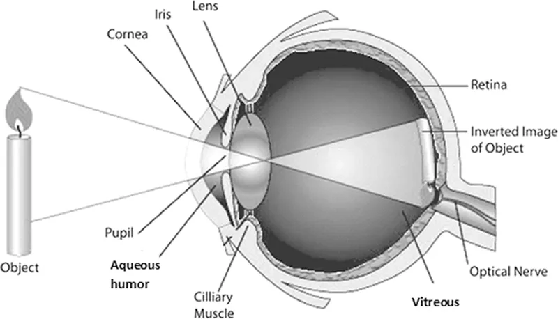
FIGURE 1.3 Image formation on the retina.
Figure 1.3 shows how the image is formed on the retina. Scattered light from the object(s) enters the eye and passes through the cornea, which bends and focuses the light on the lens. The pupil controls the amount of light that enters the eye. The lens then focuses the light onto the retina, forming an inverted image. The lens changes its shape to adjust the focus on both distant and near objects, a process known as accommodation.
Photoreceptors on the retina generate electrical signals which are sent to the brain via the optic nerve. The brain interprets the message and re-inverts the image. Hence, we see an upright image. There are two types of photoreceptors on the retina that capture the incoming light: rods and cones; these are shown in Figure 1.4.
The rods and cones are elongated retinal cells that collect the light that hits the retina. Rod photoreceptors work well in low light, provide black-and-white vision, and detect movements. Cones are responsible for color vision and work best in medium and bright light. There are three types of color-sensitive cones in the retina of the human eye, corresponding roughly to red, green, and blue detectors. The different colors are produced by various combinations of these three types of cones and their photopigments. White light is produced if the three types of cones are stimulated equally. Rods are located throughout the retina, while cones are concentrated...
Table of contents
Cover
Half Title
Title Page
Copyright Page
Table of Contents
Preface
Authors
1. Computer and Human Vision Systems
2. Digital Image Fundamentals
3. Machine Vision System Components
4. Machine Vision Applications in Quality Control
5. Digital Image Processing for Machine Vision Applications
6. Case Studies
7. Emerging Trends and Conclusion
Bibliography
Index
Frequently asked questions
Yes, you can cancel anytime from the Subscription tab in your account settings on the Perlego website. Your subscription will stay active until the end of your current billing period. Learn how to cancel your subscription
No, books cannot be downloaded as external files, such as PDFs, for use outside of Perlego. However, you can download books within the Perlego app for offline reading on mobile or tablet. Learn how to download books offline
Perlego offers two plans: Essential and Complete
Essential is ideal for learners and professionals who enjoy exploring a wide range of subjects. Access the Essential Library with 800,000+ trusted titles and best-sellers across business, personal growth, and the humanities. Includes unlimited reading time and Standard Read Aloud voice.
Complete: Perfect for advanced learners and researchers needing full, unrestricted access. Unlock 1.5M+ books across hundreds of subjects, including academic and specialized titles. The Complete Plan also includes advanced features like Premium Read Aloud and Research Assistant.
Both plans are available with monthly, semester, or annual billing cycles.
We are an online textbook subscription service, where you can get access to an entire online library for less than the price of a single book per month. With over 1.5 million books across 990+ topics, we’ve got you covered! Learn about our mission
Look out for the read-aloud symbol on your next book to see if you can listen to it. The read-aloud tool reads text aloud for you, highlighting the text as it is being read. You can pause it, speed it up and slow it down. Learn more about Read Aloud
Yes! You can use the Perlego app on both iOS and Android devices to read anytime, anywhere — even offline. Perfect for commutes or when you’re on the go. Please note we cannot support devices running on iOS 13 and Android 7 or earlier. Learn more about using the app
Yes, you can access A Guide for Machine Vision in Quality Control by Sheila Anand,L. Priya in PDF and/or ePUB format, as well as other popular books in Informatica & Ingegneria informatica. We have over 1.5 million books available in our catalogue for you to explore.