- 240 pages
- English
- ePUB (mobile friendly)
- Available on iOS & Android
eBook - ePub
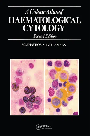
A Colour Atlas of Haematological Cytology
About this book
The second edition of this atlas has been revised and greatly en-larged to take account of recent diagnostic advances in the field of haematology. It contains 750 color photomicrographs of common and less common cell types, and is intended to supplement the per-sonal study of preparations under the microscope.
The photographs are grouped into five sections covering red cells and their precursors; nonerythroid cells of myeloid origin; lympho-cytes, plasma cells and their derivatives and precursors; miscella-neous cells of blood and marrow, including foreign cells and para-sites; and the imprint cytology of lymph nodes and spleen. Each section is preceded by a brief introduction, and an appendix pro-vides technical details of staining methods. The text accompanying the illustrations serves chiefly to identify the cells, but also gives explanations of certain unusual appearances and the cytochemical reactions where necessary.
A Colour Atlas of Haematological Cytology will be useful to students of medicine and medical laboratory science, as well as to doctors and technicians specializing in the diagnosis of haematological disease.
Trusted by 375,005 students
Access to over 1.5 million titles for a fair monthly price.
Study more efficiently using our study tools.
Information
Topic
MedicineSubtopic
HematologyPart 1
The red cells and their precursors
The nomenclature of red cell precursors is confusing. The earliest recognisable member of the red cell series, the proerythroblast, has the cytoplasmic basophilia, the nucleolated and moderately leptochromatic nucleus and the large cell size generally characteristic of primitive cells. It gives rise to a sequence of nucleated cells, the erythroblasts, which progressively develop increasingly pachychromatic nuclei, lose their nucleoli and their cytoplasmic basophilia and acquire a rising haemoglobin content. This sequence is subject to an arbitrary division into stages, the commonest division being into three:
- (1) The basophilic or early erythroblast, or normo-blast A
- (2) The polychromatic or intermediate erythroblast, or normoblast B
- (3) The orthochromatic or late erythroblast, or normoblast, or normoblast C.
There are objections to the use of many of these terms, but they are all so firmly entrenched in common usage that they must be accepted. When authors use different or more elaborate staging and nomenclature they usually define their terminology, but those who use any of the synonyms above expect them to be understood without further explanation.
The proerythroblast is not itself the functional stem cell serving as a self-maintaining progenitor of the normoblast series, but is derived from an earlier functional myeloid stem cell of unidentified morphology having pluripotential capacity for giving rise to cells of erythroid, granulocytic, monocytic and megakaryocyte-platelet lines.
Kinetic studies with radio-isotopically labelled cells suggest that four cell cycles culminating in mitoses occur during development from proerythroblast to late normo-blast, three at the proerythroblast and early basophilic normoblast stages, and the last at the polychromatic intermediate normoblast stage. Nests of erythroblasts of different stages of maturity commonly occur in apposition around a centrally situated reticulo-endothelial cell. Transfer of iron may be effected in one or other direction, and the central macrophage is often rich in stainable free iron. Late normoblasts do not undergo a further cell cycle but lose their nuclei by extrusion and give rise to marrow reticulocytes.
Reticulocytes spend up to two days in the bone-marrow before being released into the peripheral blood. There they make up normally less than 1 per cent of the red cell population and within another one to two days lose the remnants of cytoplasmic basophilia which give them their characteristic staining properties, and become orthochromatic mature red cells.
Mature red cells survive some 120 days before destruction. They are normally circular and fairly uniform in diameter, but are readily distorted by external pressure, as from neighbouring cells in a smear. Their structure as biconcave discs leads to weaker eosinophil staining at the centre than at the periphery, a feature which is least prominent at the tail of a blood smear where the cells are most spread out and flattened. In the body of the smear it becomes more conspicuo...
Table of contents
- Cover
- Title Page
- Copyright Page
- Table of Contents
- Acknowledgements
- Introduction
- Part 1 The red cells and their precursors
- Part 2 Granulocytes, monocytes and megakaryocytes
- Part 3 Lymphocytes, plasma cells and their derivatives and precursors in blood and bone marrow
- Part 4 Miscellaneous cells from bone marrow or blood smears, reticulo-endothelial cells, osteoclasts and osteoblasts, foreign cells and parasites
- Part 5 Imprints of lymph nodes and spleen; cells from pleural, ascitic and cerebrospinal fluids
- Appendix: staining techniques
- Index
Frequently asked questions
Yes, you can cancel anytime from the Subscription tab in your account settings on the Perlego website. Your subscription will stay active until the end of your current billing period. Learn how to cancel your subscription
No, books cannot be downloaded as external files, such as PDFs, for use outside of Perlego. However, you can download books within the Perlego app for offline reading on mobile or tablet. Learn how to download books offline
Perlego offers two plans: Essential and Complete
- Essential is ideal for learners and professionals who enjoy exploring a wide range of subjects. Access the Essential Library with 800,000+ trusted titles and best-sellers across business, personal growth, and the humanities. Includes unlimited reading time and Standard Read Aloud voice.
- Complete: Perfect for advanced learners and researchers needing full, unrestricted access. Unlock 1.5M+ books across hundreds of subjects, including academic and specialized titles. The Complete Plan also includes advanced features like Premium Read Aloud and Research Assistant.
We are an online textbook subscription service, where you can get access to an entire online library for less than the price of a single book per month. With over 1.5 million books across 990+ topics, we’ve got you covered! Learn about our mission
Look out for the read-aloud symbol on your next book to see if you can listen to it. The read-aloud tool reads text aloud for you, highlighting the text as it is being read. You can pause it, speed it up and slow it down. Learn more about Read Aloud
Yes! You can use the Perlego app on both iOS and Android devices to read anytime, anywhere — even offline. Perfect for commutes or when you’re on the go.
Please note we cannot support devices running on iOS 13 and Android 7 or earlier. Learn more about using the app
Please note we cannot support devices running on iOS 13 and Android 7 or earlier. Learn more about using the app
Yes, you can access A Colour Atlas of Haematological Cytology by F.G.J. Hayhoe,R.J. Flemans in PDF and/or ePUB format, as well as other popular books in Medicine & Hematology. We have over 1.5 million books available in our catalogue for you to explore.