- 742 pages
- English
- ePUB (mobile friendly)
- Available on iOS & Android
eBook - ePub
Fish Larval Physiology
About this book
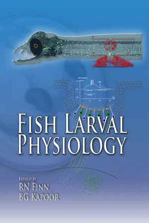
This book is intended as a resource for students and researchers interested in developmental biology and physiology and specifically addresses the larval stages of fish. Fish larvae (and fish embryos) are not small juveniles or adults. Rather they are transitionary organisms that bridge the critical gap between the singlecelled egg and sexually immature juvenile. Fish larvae represent the stage of the life cycle that is used for differentiation, feeding and distribution. The book aims at providing a single-volume treatise that explains how fish larvae develop and differentiate, how they regulate salt, water and acid-base balance, how they transport and exchange gases, acquire and utilise energy, how they sense their environment, and move in their aquatic medium, how they control and defend themselves, and finally how they grow up.
Tools to learn more effectively

Saving Books

Keyword Search

Annotating Text

Listen to it instead
Information
Part 1
Ontogeny
CHAPTER
1
Pattern Formation
Thomas E Hall
Introduction
Pattern formation during development refers to the processes by which the structure of the organism is spatially and temporally organised. On a cellular level, this requires the management of cell activities within the embryo so that a well-ordered structure develops. These processes begin prior to fertilisation, during oogenesis, and eventually result in the differentiation of the tissue and organ systems essential for homeostasis in complex organisms.
The fishes are a large and diverse group containing approximately half of known craniate species (Nelson, 1994). Early ontogeny varies substantially between clades, although this diversity is partly artifactual, since fish are a paraphyletic taxon (Collazo et al., 1994). As such, this chapter concentrates on the Actinopterygii (ray-finned fishes) and mainly on the group Teleostei, which benefits from a number of well-studied models such as the medaka (Oryzias latipes) and zebrafish (Danio rerio). In addition, the rise of intensive aquaculture in recent years has fuelled much developmental research on commercially important species.
Author’s address: Victor Chang Cardiac Research Institute, 384 Victoria Street, Darlinghurst, Sydney, NSW 2010, Australia.
E-mail: [email protected]
Early Pattern Formation
Pre-cleavage Patterning
Typically, teleosts are externally fertilising (oviparous), and the entirety of embryonic development occurs outside the mother’s body. The eggs are supplied with a large yolk volume to sustain early development in the absence of direct maternal nutrition. In the unfertilised egg, the yolk and cytoplasm are intermixed (Leung et al., 2000). The first patterning information is apparent even at this early stage as the animal/vegetal axis can be predicted on the basis of the localisation of specific maternal mRNAs (Howley & Ho, 2000). Following sperm entry and extrusion of the second polar body, the cytoplasm and yolk begin to separate, forming a single blastomere at the animal pole, sitting atop a continuous acellular yolk mass. In many species (e.g. the zebrafish; Kimmel et al., 1995) streaming of cytoplasmic constituents to the animal pole may be observed directly under the light microscope. An actin microfilament network drives this movement (Leung et al., 2000).
Cleavage
All teleosts show a discoidal meroblastic cleavage pattern, where the large yolk volume restricts cell division to a small area at the animal pole. The early embryo forms as a small disc sitting atop the large yolk. Holoblastic cleavage, where division splits the entire mass, is thought to be the ancestral condition, and is common in many invertebrate chordate phyla, as well as the lampreys. However, it appears that most animals with telolecithal eggs (those with a high ratio of yolk:cytoplasm) have evolved the meroblastic “shortcut”. A meroblastic cleavage pattern allows cell division to proceed rapidly, and avoids the need for synthesis of large amounts of new cell membrane. Indeed, meroblastic cleavage has evolved independently at least five times within the craniates (once in each of the lineages leading to the hagfish, elasmobranchs, coelacanths, teleosts and amniotes; Collazo et al., 1994). Interestingly, present-day mammals show a reversion to holoblastic cleavage, possibly as a result of gaining nutrition directly from the mother, rather than having to lay down copious quantities of yolk.
The first divisions are vertical and there is no cytoplasmic growth during this period, resulting in a decrease in blastomere size with each successive division. Early cleavages tend to be incomplete, and the initial blastomeres maintain cytoplasmic bridges with the yolk cell. Cleavages occur synchronously between embryos and the first cell cycles are easy to follow within clutches. Mitotic spindles can often be seen as the disc cleaves to form two distinct cells (Kimmel et al., 1995; Hall et al., 2004). The second cleavage is also horizontal and results in a 2×2 array. Timing of the cell cycle during the first cleavage events is highly synchronous, with each cycle having approximately the same length (Marrable, 1965; Kimmel et al., 1995). Around the 16 to 32 cell stage however, the mass of cells (the blastodisc), becomes stratified into more than one layer. In most species the blastomeres are regular in size and shape. However, inter- and intra-specific variation exists on the general pattern. In the Atlantic cod (Gadus morhua) horizontal stratification of the cell mass usually occurs at the sixth cleavage, between the 32 and the 64 cell stages, but is frequently observed at the previous or following cleavage (Hall et al., 2004). In the medaka, the first horizontal cleavage is the fifth, occurring between the 16 and 32 cell stages (Iwamatsu, 1994) and in the ice goby (Leucopsarion petersii) it occurs even earlier, between the 4 and 8 cell stages (Nakatsuji et al., 1997). Unusually within the teleosts, eggs of the wolffish (Anarhichas lupus), have unequal blastomere sizes during early divisions (Pavlov et al., 1992). During later cleavages it is only the marginal blastomeres (sometimes called Wilson cells), positioned on the periphery of the cell mass (now called the blastodisc), which maintain their cytoplasmic bridges with the yolk.
The Early Blastula and the Yolk Syncytial Layer
Loss of cytoplasmic bridges coincides with the formation of the yolk syncytial layer (YSL; Trinkaus, 1992). Around the ninth and tenth cleavages, the marginal blastomeres collapse into a single large, multinucleate (or syncytial) cell (Kimmel & Law, 1985). The YSL is an organ unique to teleosts that no longer undergoes cytoplasmic cleavage. The nuclei, however, continue to divide mitotically, and spindles may be seen in the cytoplasm as the YSL spreads beneath the blastodisc, separating the true blastomeres from the yolk. Around the 12th cleavage the YSL nuclei become post-mitotic (Kane et al., 1992). Three cellular layers are now visible in the blastula; an enveloping layer (EVL) constituting the most external cells, which is epithelial in nature and covers the blastoderm; a middle layer of deep cells which will become the embryo proper, and the YSL lying between the deep cells and the yolk. The YSL is continuous with the yolk cytoplasmic layer (YCL), a thin layer of anuclear cytoplasm surrounding the yolk. The YSL itself is often described as possessing two domains, the internal YSL, which spreads beneath the blastodisc, and the external YSL which lies around the circumference. Tight junctions between the YSL and the overlying blastomeres mark the boundary between the internal and external domains. The thinner internal YSL (beneath the blastodisc) is populated by nuclei derived from the external YSL (Kimmel & Law, 1985).
In the early development of many animals, the initial rapid cleavage stages are followed by a period where the cell cycle lengthens, the embryo begins to transcribe its own RNA, and the cells of the blastula become motile, in preparation for gastrulation. This is termed the mid-blastula transition (MBT; Kane & Kimmel, 1993). The teleost MBT begins around cell cycle eight-ten (cycle ten in the zebrafish and the Atlantic cod). From around the seventh cleavage, the cells begin to show metasynchrony in their divisions, and within a further two or three cell cycles, cleavage events cannot be distinguished. In contrast to the early cleavages when there was little net cell movement, cell divisions now often spread daughter cells more than a cell diameter apart, causing a mild dispersion of progeny and intermingling of distant cell lineages (Kimmel & Law, 1985). Pseudopodia are also present, consistent with the activation of cell motility. However, the fate map in the early blastula is non-determinative; the progeny of any blastomere may contribute to all tissues in the embryo. Cell movements at this stage are not “directed or coherent” (Kane & Adams, 2002). Interestingly, the onset of ...
Table of contents
- Cover
- Half Title
- Title Page
- Copyright Page
- Preface
- Table of Contents
- List of Contributors
- Part 1: Ontogeny
- Part 2: Respiration & Homeostasis
- Part 3: Nutrition and Energy
- Part 4: Sensory Physiology
- Part 5: Movement
- Part 6: Control and Defense
- Part 7: Functional Changes in Form
- Glossary
- Species Index
- Common Name Index
- Subject Index
Frequently asked questions
Yes, you can cancel anytime from the Subscription tab in your account settings on the Perlego website. Your subscription will stay active until the end of your current billing period. Learn how to cancel your subscription
No, books cannot be downloaded as external files, such as PDFs, for use outside of Perlego. However, you can download books within the Perlego app for offline reading on mobile or tablet. Learn how to download books offline
Perlego offers two plans: Essential and Complete
- Essential is ideal for learners and professionals who enjoy exploring a wide range of subjects. Access the Essential Library with 800,000+ trusted titles and best-sellers across business, personal growth, and the humanities. Includes unlimited reading time and Standard Read Aloud voice.
- Complete: Perfect for advanced learners and researchers needing full, unrestricted access. Unlock 1.4M+ books across hundreds of subjects, including academic and specialized titles. The Complete Plan also includes advanced features like Premium Read Aloud and Research Assistant.
We are an online textbook subscription service, where you can get access to an entire online library for less than the price of a single book per month. With over 1 million books across 990+ topics, we’ve got you covered! Learn about our mission
Look out for the read-aloud symbol on your next book to see if you can listen to it. The read-aloud tool reads text aloud for you, highlighting the text as it is being read. You can pause it, speed it up and slow it down. Learn more about Read Aloud
Yes! You can use the Perlego app on both iOS and Android devices to read anytime, anywhere — even offline. Perfect for commutes or when you’re on the go.
Please note we cannot support devices running on iOS 13 and Android 7 or earlier. Learn more about using the app
Please note we cannot support devices running on iOS 13 and Android 7 or earlier. Learn more about using the app
Yes, you can access Fish Larval Physiology by Roderick Nigel Finn in PDF and/or ePUB format, as well as other popular books in Biowissenschaften & Physiologie. We have over one million books available in our catalogue for you to explore.