Originally published in 1981, this volume represents the edited proceedings of the third symposium on eye movements and behaviour sponsored by the US Army Human Engineering Laboratory. The conference, titled "The Last Whole Earth Eye Movement Conference" was held in Florida in February 1980. As the conference approached, seizure of the American hostages by the Iranian militants, the Russian invasion of Afghanistan, and the uncertain economic outlook around the world made it appear as though the title was a self-fulfilling prophecy. But the meeting proved highly successful and people throughout the world seemed to be adapting to the stresses of international tension, making the possibility of subsequent meetings more likely. The present volume is intended to serve as a complementary text to the earlier texts Eye Movements and Psychological Processes (Monty & Senders, 1976) and Eye Movements and the Higher Psychological Functions (Senders, Fisher & Monty, 1978), rather than a revision and update of them.

eBook - ePub
Eye Movements
Cognition and Visual Perception
- 372 pages
- English
- ePUB (mobile friendly)
- Available on iOS & Android
eBook - ePub
Eye Movements
Cognition and Visual Perception
About this book
Trusted by 375,005 students
Access to over 1 million titles for a fair monthly price.
Study more efficiently using our study tools.
Information
I Infant and Developing Mechanisms
Saccades, smooth pursuit, and optokinetic nystagmus— when do they begin? Is the ability to perform the various eye movements innate, or must their development correspond to changes in sensitivity of the visual system and the maturation of the visual cortex? What are the implications of the developing eye movement system for perceiving form, adapting to moving stimuli, and perceiving eccentric stimuli? Are there methodological problems in matching the measurement device to the infants' capability of being a cooperative participant? These are some of the questions of concern that are addressed in Part I.
Hainline, in Chapter I. 1 examines the form perception of infants and finds that there is significant variability between infants' scanning patterns. Some infants show very narrow or restrictive patterns, while others show broad scanning patterns with little regularity, irrespective of the size of the form viewed. Procedural implications weigh heavily in her research, both in the handling of infant methodologically and as related to past research findings.
In Chapter 1.2, Maurer and Lewis take issue with the traditional perceptual learning theorists notion that the shape of a stimulus can be resolved only after a thorough scanning, by presenting infants with various "interesting" patterns 20° or 30° in the periphery. Although there may be some question about the infants' selectivity as far out as 30°, lesser eccentricities prove compelling stimuli to the infants except when shown in the presence of competing centrally viewed stimuli. Further, it is their contention that decisions about peripheral stimuli can be made as early as 3 months and these are purely perceptual without cognitive interference. Maurer and Lewis also express their concern over the high degree of poor "voluntary" participation of these infants, generally concluding that they make bad subjects.
In Chapter 1.3, Aslin reports on a highly successful monitoring technique in which infants from 3 weeks to 4 months old are tested for the presence and efficiency of smooth pursuit movements, one of the so-called more primitive eye movements. He examines various aspects of the smooth pursuit system by age of emergence, tracking accuracy of various velocities, possible evidence for predictive tracking, and any evidence for a velocity gain mechanism in this smooth pursuit system independent of saccades. While providing many answers to these concerns, the overriding issue of an independent smooth pursuit movement system receives confirmation.
In the final chapter of this section, Atkinson and Braddick describe the development of optokinetic nystagmus in infants as highly correlated with cortical binocularity. In the four experiments described, OKN is related to development, abnormal visual development i.e. strabismus and amblyopia, and to its relationship with other systems like smooth pursuit. For infants under 3 months of age, an asymmetry occurs for nasal-temporal and temporal-nasal direction of the stimulus motion for elicitation of OKN. This asymmetry generally seems to dissipate as the infants grow older. Qualifications were made also on short and long OKN, but a general independence of the OKN system and smooth pursuit system seems confirmed. Somewhat cautiously, these data are interpreted as suggesting that binocular cortex is a necessary condition for symmetrical OKN, but there is as yet little direct evidence that the development of cortical binocularity is the critical factor for the onset of symmetrical OKN in infancy.
I.1 Eye Movements and Form Perception in Human Infants
Louise Hainline
Brooklyn College of C.U.N.Y.
Brooklyn College of C.U.N.Y.
Infancy stands at the beginning of a long course of psychological development. Human infants are faced with the task of appreciating a complex environment with what appears to be a limited set of resources. Vision is obviously one of these resources and infants can be observed actively scanning their environment with organized sequences of eye movements and fixations within minutes after birth. Visual information is an important factor in cognitive and social development, as evidenced in studies of the concept of the permanence of objects and attachment to a caretaker (Fraiberg, 1977). However, an understanding of perceptual development at the beginning of life may also contribute to an understanding of perceptual processes at other stages in the life span. For example, one might study visual development as a means of addressing hoary theoretical questions about the nature of perception as expressed in the polarization between a Gestalt position on innate organization versus a "constructionist" approach supporting the role of learning and experience in perception.
For many years the infant was regarded as a largely reflexive creature at the mercy of environmental exigencies. Infants probably haven't changed significantly in the past decade, but developmental psychology's conception of them has. New research on infant psychological processes has generated a revised view of the competent infant, almost hungrily grappling with and trying to master the environment. To some extent, this change in perspective has resulted from methodological innovation and technological advances. Even brief exposures to infants allow the observation of characteristics that make them poor candidates for subjects in psychological research. Evanescent states of wakefulness, unbridled emotions, and periodic dampness pose real challenges to the researcher, who has needed a battery of behavioral and psychophysical techniques to make headway.
Since the pioneering work of Fantz (1956, 1958), it has been reasoned that one way of learning what the infant sees is to study what he looks at. This line of reasoning has led from studies of visual choice behavior (the so-called visual preference technique) to increasing sophistication in the measurement of actual eye movement patterns. Early techniques developed by Salapatek and Kessen (1966), Salapatek (1968), and Haith (1978) were based on corneal reflection photography, were painfully slow, and involved various techniques for hand scoring characteristics of the scanning eye "frozen" for analysis one to four times a second. Calibration data on the accuracy of the technique were difficult if not impossible to obtain, but allowed relatively precise specifications of eye position during scanning.
System Detail
The research technique used in my laboratory is based on the principles of this earlier work on infant eye movement measurement, but allows greater speed and potentially greater accuracy. The system is based on a low light level television system originally designed for measuring adult eye movements, a Gulf + Western, Applied Sciences Laboratory (ASL) Model 1994 Eye View Monitor. Extensive modifications to the apparatus have been made for work with infants, and, in view of its unique nature, it will be described in more detail, although the basic technique is a familiar one. The system's principle of operation is show schematically in Fig. 1. It has a low light level invisible infrared source whose light is collimated so that rays are parallel when they reach the subject's eye, after reflection by an IR reflecting, visible transmitting dichroic mirror. A small amount of the light that enters the pupil is reflected by the retina, and is photographed by a very sensitive television camera. The camera provides an image of the eye with a bright pupil enlarged on a monitor. Actually, only part of the incident light enters the eye; a fraction is reflected by the cornea and appears on the television image as a small bright virtual image, the corneal reflection or first Purkinje image, superimposed on the bright pupil. As the eye rotates to look at a portion of the visual field, the corneal reflection moves differentially with respect to the pupil, but if the entire head moves, within limits, the pupil and corneal reflection move together. Thus if the position of the head is stabilized, analysis of shifts of the corneal reflection relative to the pupil can specify direction of regard. The ASL Eye View Monitor automates this estimation so current eye position is available 60 times a second.
During testing, the infant is held against the shoulder of an experimenter who gently stabilizes the infant's head; the infant looks through the dichroic beam splitter at a rear projection screen on which the stimuli appear. Figure 2 illustrates more clearly the other elements of the system. The "eye camera" as described forms an image of the corneal reflection and the pupil. The "face camera" provides the experimenter with a picture of the infant's face, and so permits the
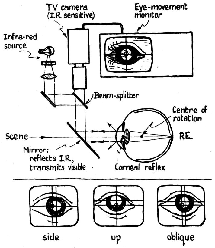
FIG. 1. A schematic representation of the essential elements and principle of operation of a television based infrared eye movement recording system. The lower portion of the figure illustrates the differential movement of the corneal reflex with respect to the center of the pupil during eye movements that is the basis for the calculation of eye position.
proper positioning of the eye. The experimenter cannot see the stimuli and so bias any of the responses. The "scene camera" photographs the stimulus viewed by the infant. Outputs of both the scene and eye cameras are fed into the eye movement monitor, which calculates direction of the infant's regard every 60th of a second and relates it to the stimulus. Data on eye position are fed into a minicomputer that provides some on-line analysis, data storage, and experimental control. The system minimizes subjective decisions by human scorers and gives a large amount of data even for brief stimulus presentations. The digitized eye movement records are subsequently plotted as patterns of successive fixations on the stimulus and are subjected to other computer analyses.
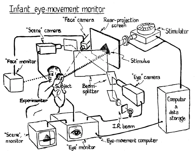
FIG. 2. A schematic representation of the elements of an infrared eye movement recording system for use with infants.
If relative eye movement information is desired as in measuring responses like optokinetic nystagmus, this system can achieve an accuracy of under .5° with infants, but for many research questions, like those involving pattern perception, data on absolute position in space are required. When using older subjects, they can be told to fixate a few specifiable points in the field, and calibration is accomplished by adjusting gains on the apparatus or performing mathematical corrections on the data. True fixation could differ from obtained estimates of fixation for several functionally distinct reasons—for example, instrument error, corneal irregularities, or because the fovea is not actually on the eye's optic axis, which intersects the center of the pupil which is used as the reference point for eye position calculations. The technique further assumes that the fovea is the most relevant portion of the retina for information about the point of regard; for young infants, data on retinal development suggest this may be an improper assumption (Mann, 1964). To estimate the extent of the sum of these errors in infants, we have developed techniques for calibrating the eye movements of subjects who cannot be instructed to fixate points A, B, and C in sequence.
The Calibration Algorithm
Since the attention span of infants is notoriously brief, our strategy has been to collect scanning information to the stimuli of interest first, and then, if the infant is still cooperative, to move into the calibration procedure. The calibration results are later used to adjust the fixation estimates. A sequence of small flashing lights is presented one at a time to the subjects. The lights are at known coordinates in the same system as the fixation data. An experimenter observes the output on the scene monitor and signals the computer to collect data when the infant appears to be fixating the reference light. The logic of the routine ultimately involves the provision of some "average" point of fixation for each reference light, but the routine is flexible so that each set of data potentially involving fixation of the reference point may also involve non-fixation data representing "wobble" in an attempt to maintain a steady fixation (which would probably be symmetric about a "true mean") or systematic movement toward or away from the reference point (which would probably not be symmetrical about the true mean). The calibration algorithm first estimates fixation coordinates for each reference point by calculating, separately for X and Y, a mean and variance of all the points in the set. Any points lying be...
Table of contents
- Cover
- Half Title
- Title
- Copyright
- Original Title
- Original Copyright
- Contents
- Participants and Contributors
- Preface
- PART I: INFANT AND DEVELOPING MECHANISMS
- PART II: ILLUSIONS AND AFTEREFFECTS
- PART III: PICTURES AND PICTORIAL PROCESSING
- PART IV: THEORY AND METHOD
- PART V: SEARCH AND SCANNING
- PART VI: CAN EYE MOVEMENTS SAVE THE EARTH?
- PART VII: REFERENCES
- Author Index
- Subject Index
Frequently asked questions
Yes, you can cancel anytime from the Subscription tab in your account settings on the Perlego website. Your subscription will stay active until the end of your current billing period. Learn how to cancel your subscription
No, books cannot be downloaded as external files, such as PDFs, for use outside of Perlego. However, you can download books within the Perlego app for offline reading on mobile or tablet. Learn how to download books offline
Perlego offers two plans: Essential and Complete
- Essential is ideal for learners and professionals who enjoy exploring a wide range of subjects. Access the Essential Library with 800,000+ trusted titles and best-sellers across business, personal growth, and the humanities. Includes unlimited reading time and Standard Read Aloud voice.
- Complete: Perfect for advanced learners and researchers needing full, unrestricted access. Unlock 1.4M+ books across hundreds of subjects, including academic and specialized titles. The Complete Plan also includes advanced features like Premium Read Aloud and Research Assistant.
We are an online textbook subscription service, where you can get access to an entire online library for less than the price of a single book per month. With over 1 million books across 990+ topics, we’ve got you covered! Learn about our mission
Look out for the read-aloud symbol on your next book to see if you can listen to it. The read-aloud tool reads text aloud for you, highlighting the text as it is being read. You can pause it, speed it up and slow it down. Learn more about Read Aloud
Yes! You can use the Perlego app on both iOS and Android devices to read anytime, anywhere — even offline. Perfect for commutes or when you’re on the go.
Please note we cannot support devices running on iOS 13 and Android 7 or earlier. Learn more about using the app
Please note we cannot support devices running on iOS 13 and Android 7 or earlier. Learn more about using the app
Yes, you can access Eye Movements by Dennis F. Fisher, Richard A. Monty, John W. Senders, Dennis F. Fisher,Richard A. Monty,John W. Senders in PDF and/or ePUB format, as well as other popular books in Psychology & Cognitive Psychology & Cognition. We have over one million books available in our catalogue for you to explore.