- 264 pages
- English
- ePUB (mobile friendly)
- Available on iOS & Android
eBook - ePub
Atlas of Feline Anatomy For Veterinarians
About this book
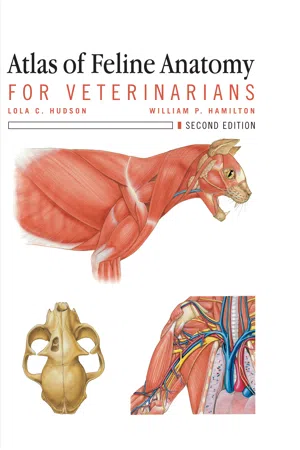
Presenting more than 266 full color anatomic drawings arranged by organ system, this book is dedicated exclusively to feline anatomy with emphasis on those areas of anatomy that are frequently encountered in clinical practice. It includes a highly detailed chapter on special senses which collects and organizes difficult to find information for quick access. Nomenclature is from Nomina Anitomica Veterinaria so that the feline anatomy is in line with that used in other textbooks of veterinary anatomy of the domestic animals. The book accurately captures the anatomy pertinent to clinical veterinary medicine.
Trusted by 375,005 students
Access to over 1.5 million titles for a fair monthly price.
Study more efficiently using our study tools.
Information
Topic
MedicineSubtopic
Veterinary MedicineContents
Chapter One General Information and Physical Examination
Lola Hudson
Chapter Two Integumentary System
Cherie Pucheu-Haston
Chapter Three Musculoskeletal System
Lola Hudson and William Hamilton
Chapter Four Cardiovascular System
Jill Barnes
Chapter Five Lymphoid System
Mary Tompkins and Kristina Howard
Chapter Six Endocrine System
Antonella Borgatti Jeffreys and David Waters
Chapter Seven Respiratory System
Bonnie Smith
Chapter Eight Digestive System
Jill Barnes
Chapter Nine The Urogenital System
Bonnie Smith
Chapter Ten Nervous System
Lola Hudson
Chapter Eleven Special Sensory Organs
Lola Hudson
Index
Chapter 1
General Information and Physical Examination
General Information
The “house” cat was fully domesticated at least by 1600 BC, after domestication of the dog. While a cat skeleton from some 9500 years ago was discovered in Cyprus, this was believed to be during the period of domestication. Most authorities cite Egypt as a location for domestication but a more exact place is not known. It is believed that domestication coincided with the invention of grain silos, and that the human need for rodent control and the cat’s need for a ready food source resulted in a mutually beneficial, but still aloof, relationship.
The cat was deified in ancient Egypt, being worshipped as Bastet, the goddess of fertility - both human and agricultural. This special esteem of cats resulted in attempts to prevent their export from Egypt, but eventually domestic cats appeared in other areas of the Mediterranean. The domesticated cat is now found throughout the populated world and over 35 breeds have been developed, although not all breeds are recognized by all groups.
The Cat Fanciers Association currently recognizes 37 breeds. An abbreviated list of breeds ( Table 1-1) is included in this chapter.
The domestic cat has the scientific name of Felis catus (F. cattus), but has also been identified under the name Felis domes- ticus (F. domestica), as the taxonomy underwent changes in 1996. Cats belong to the family Felidae, which includes the “big cats,” to the order Carnivora, and to the class Mammalia. Overall, the cat shows less variation in body size and skull shape than is seen among dogs, which suggests that there is a more uniform genetic makeup in cats. Nevertheless, relatively long-faced cats (Siamese), short-faced cats (Persian) and medium-faced cats (domestic short hair) are found among the various recognized breeds.
Table 1-1
Abbreviated List of Recognized Feline Breeds in the United States
Abbreviated List of Recognized Feline Breeds in the United States
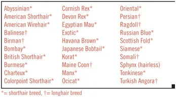
* = shorthair breed, f= longhair breed
The pet cat population in the United States has steadily increased over the last decade. The American Veterinary Medical Association reports that there are more cats than dogs in USA households as the companion animal. Such numbers have influenced the interest of the veterinary profession as seen by the increase in numbers of feline-related seminars at various local, regional, and national professional meetings. This interest has also lead to a boom in textbooks and monographs on feline medicine and surgery for the veterinary profession or inclusion of more feline-related material in “small animal” textbooks.
Physical Examination
A good, thorough physical examination takes only a few minutes to perform but reveals a wealth of information to the veterinarian. In order to be efficient, the routine examination is performed systematically, concentrating on the same things in the same order. The precise order is not important as long as it is done the same way each time. The procedure then becomes second nature and the likelihood of overlooking something is minimized. As many of the examination procedures are irritating to many cats, becoming practiced at a technique is important. A large portion of a physical examination is based on palpation. Which structures are palpable in a normal cat is dependent upon the body condition of the cat and the skill and experience of the veterinarian. The following is one method of performing the physical examination.
First, the cat is removed from its carrier. If it can move about without escaping, the cat is observed walking, noting the mentation, posture and gait for balance and control, as well as its general attitude. Once the cat is placed on an examination table, the hair coat is checked for indications of self-grooming, for areas of alopecia and for ectoparasites.
Next, the head becomes the focus. The dorsum of the skull is palpated while noting the health of the hair and skin. Both eyes are examined for size, shape, and position. The upper eyelid can be retracted to observe the sclera and conjunctiva for color. The presence of a normal tear film is noted by the glistening appearance of the cornea. Using the upper eyelid for protection, each orb is gently pressed to roughly compare intraocular pressure. (This step can be substituted with the use of ocular pressure measuring devices such as a Tonopen®.) During this procedure, the third eyelid may move into clear view and be assessed. The corneas, irides, and pupils are checked for smoothness, clarity, and symmetry. With a strong light source such as a transilluminator, the pupillary light reflex of each eye is assessed. The clarity and uniformity of the lens is evaluated. The fundus is examined taking care to visualize the retinal blood vessels, tapetum lucidum, non-tapetal areas and the optic disc. Both lateral and medial commissures are gently touched to observe the palpebral (blink) reflex, assessing cranial nerve V (sensory) and cranial nerve VII (motor) function. Care is taken not to stimulate the tactile hairs or induce a visual menace response instead of a palpebral reflex by using the tips of closed forceps brought over the head, caudal to rostral. A menace response is elicited by a small, quick hand movement toward each eye or an up and down motion in front of the eye while blocking the view of the other eye. Care is taken not to stimulate any tactile hairs with either the hand or by air movement.
The ears are assessed for normal upright position. The pinnae are examined on the concave and convex surfaces, and the canals are checked for abnormalities. An otoscopic examination of the external auditory canal and tympanic membrane is performed.The hairs inside the pinna are gently touched to elicit a twitch assessing cranial nerve VII motor function to auricular muscles.
The skin and hair coat of the nose are examined. The paranasal sinuses and recesses of the skull are assessed for sensitivity by pressing firmly dorsal to each eye, ventral to each eye, and on each side of the nose. Both nares are examined for symmetry and discharge. Gently touching just inside the nares with closed, blunt hemostats elicits a strong aversive reaction, assessing cranial nerve V sensory function.
The hair and skin of the lips are checked as well as the tactile hairs. The symmetry of the lips is noted. Lifting the lips allows examination of the gums, including color, and teeth. Capillary refill time can be ascertained by pressing the gum with a fingertip to blanch out the blood and observing how quickly the area turns pink again. Gently opening the mouth affords a quick assessment of the teeth, tongue, to...
Table of contents
- Cover
- Title
- Copyright
- Table of Contents
Frequently asked questions
Yes, you can cancel anytime from the Subscription tab in your account settings on the Perlego website. Your subscription will stay active until the end of your current billing period. Learn how to cancel your subscription
No, books cannot be downloaded as external files, such as PDFs, for use outside of Perlego. However, you can download books within the Perlego app for offline reading on mobile or tablet. Learn how to download books offline
Perlego offers two plans: Essential and Complete
- Essential is ideal for learners and professionals who enjoy exploring a wide range of subjects. Access the Essential Library with 800,000+ trusted titles and best-sellers across business, personal growth, and the humanities. Includes unlimited reading time and Standard Read Aloud voice.
- Complete: Perfect for advanced learners and researchers needing full, unrestricted access. Unlock 1.5M+ books across hundreds of subjects, including academic and specialized titles. The Complete Plan also includes advanced features like Premium Read Aloud and Research Assistant.
We are an online textbook subscription service, where you can get access to an entire online library for less than the price of a single book per month. With over 1.5 million books across 990+ topics, we’ve got you covered! Learn about our mission
Look out for the read-aloud symbol on your next book to see if you can listen to it. The read-aloud tool reads text aloud for you, highlighting the text as it is being read. You can pause it, speed it up and slow it down. Learn more about Read Aloud
Yes! You can use the Perlego app on both iOS and Android devices to read anytime, anywhere — even offline. Perfect for commutes or when you’re on the go.
Please note we cannot support devices running on iOS 13 and Android 7 or earlier. Learn more about using the app
Please note we cannot support devices running on iOS 13 and Android 7 or earlier. Learn more about using the app
Yes, you can access Atlas of Feline Anatomy For Veterinarians by Lola Hudson,William Hamilton in PDF and/or ePUB format, as well as other popular books in Medicine & Veterinary Medicine. We have over 1.5 million books available in our catalogue for you to explore.