The aim of this chapter is to provide a detailed and practical introduction to immunoassays and set the overall context for the other detailed chapters on specific key elements of immunoassays. The production of antibodies, various antibody structures and their fundamental role in immunoassays is outlined. Comprehensive guides to different immunoassay formats ranging from direct to competitive are provided, and subsequently, important signalling systems, including colourimetric and fluorescence-based approaches, are examined. Finally, electrical, mechanical and optical signal transduction mechanisms that are used in the next generation of immunoassays are described.
1.1 Introduction to Antibodies and Immunoassays
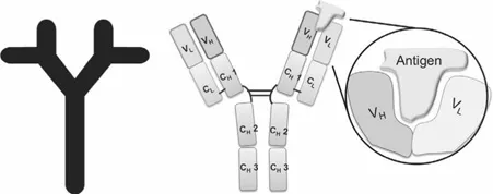
Immunoassays are biochemical tests that utilise immunoglobulins (Ig) (antibodies) as high sensitivity binders to detect the presence of molecules that are present at low concentrations. Antibodies are an integral part of the humoral immune system and are nature’s major recognition devices. They are found on the surface of B-cells (B-lymphocytes) and recognise the presence of foreign antigens. The B-cell can detect and signal the presence of an extensive range of antigens, consisting of molecules ranging from prions to viruses, drugs, toxins and aberrant biomolecules that invade our bodies. Immunoglobulin proteins exist in many different formats, the five main isotypes of immunoglobulin are IgG (shown in Fig. 1.1), IgM, IgD, IgA and IgE.
Immunoassays: Development, Applications and Future Trends
Edited by Richard O’Kennedy and Caroline Murphy
Copyright © 2017 Pan Stanford Publishing Pte. Ltd.
ISBN 978-981-4669-97-9 (Hardcover), 978-1-315-20654-7 (eBook)
www.panstanford.com
Antibodies are at the forefront of targeted therapeutics and diagnostics due to their natural high affinities and excellent half-lives. They can be readily manipulated using standard molecular biological techniques into customised antibodies that are tailored to perform efficiently in their chosen end-point application. The biopharmaceutical industry has heavily invested in antibody-based diagnostics and therapeutics, and the latter currently represents the largest and fastest growing class of biopharmaceuticals [1, 2].
Antibodies are generally represented in three forms: (i) poly-clonal (produced from a mixture of various B-cell clones), (ii) monoclonal (secreted from a single clone of B-cells) and (iii) recombinant antibodies (the product of the genetic manipulation of antibody genes) [3, 4].
1.1.1 Polyclonal Antibody Production
Polyclonal antibody production involves the immunisation of animals with an antigen and adjuvant (immune system stimulating material) mixture, resulting in the activation of multiple B-cells targeting different antigen epitopes. This produces a vast number of antibodies with different specificities and epitope affinities [3]. These polyclonal antibodies are purified from the serum of immunised animals. Polyclonal antisera can be obtained relatively quickly (6–12 weeks for highly immunogenic antigens) but the time may be antigen-dependent. Methods of purification are generally associated with the antibody type and the intended application(s) for the antibody. These are listed in Table 1.1 [5] and are discussed in detail in Chapter 12.
Several key steps need to be considered for the production of polyclonal antibodies, including (i) preparation/availability of antigen, (ii) choice of host species, (iii) injection regime, (iv) monitoring antibody response to immunogen and (v) collection/purification of antibodies. The choice of host is of particular importance when developing polyclonal antibodies against human derived targets, as a large number of proteins are highly conserved throughout mammalian evolution, and, are, therefore, common to many mammalian species. Hence, immunisation of such proteins into rabbits and mice, may generate a limited immune response [6]. The use of a species more phylogenetically distant from humans such as chickens (that diverged from mammalian genomes some 310 million years ago [7]) is an ideal alternative for immunisation and selection of antibodies against highly conserved human proteins [6, 8].
Table 1.1 Polyclonal antibody purifiation methods [5]
Purification type | Methodologies involved in the purification |
Crude | Precipitation of a subset of total serum proteins that includes immunoglobulins. |
General | Affinity purification of certain antibody classes (e.g. IgG). |
Specific | Affinity purification of only those antibodies in a sample that bind to a particular antigen molecule. |
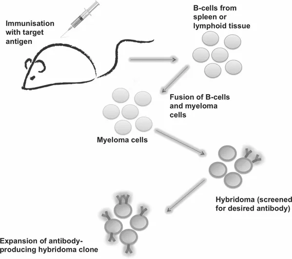
1.1.2 Production of Monoclonal Antibodies
The development of ‘hybridoma technology’ won Georges Köhler and César Milstein the Nobel Prize in Physiology or Medicine in 1984. They created an immortal cell line capable of producing an endless supply of identical antibodies with known specificity called ‘monoclonal antibodies’ signifying the fact that they were derived from a single hybrid cell [9]. The production of monoclonal antibodies is shown in Fig. 1.2 [3].
1.1.3 Recombinant Antibody Fragments
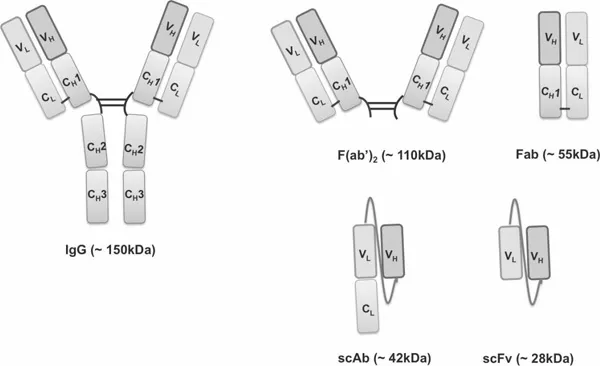
Recombinant antibodies are highly attractive for customised antibody development as genetic manipulations can be easily employed to precisely tailor their specificity and biophysical properties [10–14]. The ability to readily generate bulk quantities of recombinant proteins, with low production costs in Escherichia coli, has fuelled the emergence of an assortment of distinct antibody constructs that can be employed in a wide variety of applications. Some common recombinant antibody constructs are shown in Fig. 1.3.
The smallest antibody fragment that retains the full monovalent antigen binding capabilities of a human IgG is the variable fragment (Fv) and it is comprised of the variable heavy (VH) and variable light (VL) domains held together by non-covalent interactions [12, 13]. The single chain fragment variable (scFv) is more stable than the Fv (which is more prone to aggregation than the scFv) and typically, a flexible 15 residue glycine/serine ((Gly4Ser)3) linker is incorporated to link the chains together and aid antibody folding and stability during bacterial expression [14].
The antigen-binding fragment (Fab) is a larger, more stable fragment of approximately 50 kDa. The Fab fragment consists of the variable heavy, variable light, constant heavy and constant light chains (VH & CH1 and VL & CL). An interchain disulphide bond exists between the CH1 and CL (Fig. 1.3) [15].
There are various advantages and disadvantages associated with the different types of recombinant antibody structures that can be engineered. It should also be noted that antibody fragments can be relatively easily reformatted for improved characteristics such as expression [16], reduction in non-specific binding [17] and enhanced specificity [18]. The selection of superlative antigen-specific recombinant fragments can be achieved using a process known as phage display technology.
1.1.3.1 Production of recombinant antibodies by phage display technology
When George P. Smith first demonstrated in 1985, that the link between phenotype and genotype could be established in filamentous bacteriophage, the advent of phage display technology emerged [19]. Over the last few decades an increased understanding of antibody structure and function has allowed phage display to become one of the most powerful tools in the controlled selection ...