eBook - ePub
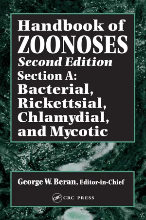
Handbook of Zoonoses, Second Edition, Section A
Bacterial, Rickettsial, Chlamydial, and Mycotic Zoonoses
- 560 pages
- English
- ePUB (mobile friendly)
- Available on iOS & Android
eBook - ePub
Handbook of Zoonoses, Second Edition, Section A
Bacterial, Rickettsial, Chlamydial, and Mycotic Zoonoses
About this book
This multivolume handbook presents the most authoritative and comprehensive reference work on major zoonoses of the world. The Handbook of Zoonoses covers most diseases communicable to humans, as well as those diseases common to both animals and humans. It identifies animal diseases that are host specific and reviews the effects of various human diseases on animals. Discussions address diseases that remain important public and animal health problems and the techniques that can control and prevent them.
The chapters are written by internationally recognized scientists in their respective areas of disease, who work or have worked extensively in the most affected areas of the world. The emphasis for each zoonosis is on the epidemiology of the disease, the clinical syndromes and carrier states in infected animals and humans, and the most current methods for diagnosis and approaches to control. For infectious agents or biologic toxins, which may be transmitted by foods of animal origin, a strong focus is placed on food safety measures. The etiologic and therapeutic aspects of each disease important to epidemiology and control are identified.
Frequently asked questions
Yes, you can cancel anytime from the Subscription tab in your account settings on the Perlego website. Your subscription will stay active until the end of your current billing period. Learn how to cancel your subscription.
No, books cannot be downloaded as external files, such as PDFs, for use outside of Perlego. However, you can download books within the Perlego app for offline reading on mobile or tablet. Learn more here.
Perlego offers two plans: Essential and Complete
- Essential is ideal for learners and professionals who enjoy exploring a wide range of subjects. Access the Essential Library with 800,000+ trusted titles and best-sellers across business, personal growth, and the humanities. Includes unlimited reading time and Standard Read Aloud voice.
- Complete: Perfect for advanced learners and researchers needing full, unrestricted access. Unlock 1.4M+ books across hundreds of subjects, including academic and specialized titles. The Complete Plan also includes advanced features like Premium Read Aloud and Research Assistant.
We are an online textbook subscription service, where you can get access to an entire online library for less than the price of a single book per month. With over 1 million books across 1000+ topics, we’ve got you covered! Learn more here.
Look out for the read-aloud symbol on your next book to see if you can listen to it. The read-aloud tool reads text aloud for you, highlighting the text as it is being read. You can pause it, speed it up and slow it down. Learn more here.
Yes! You can use the Perlego app on both iOS or Android devices to read anytime, anywhere — even offline. Perfect for commutes or when you’re on the go.
Please note we cannot support devices running on iOS 13 and Android 7 or earlier. Learn more about using the app.
Please note we cannot support devices running on iOS 13 and Android 7 or earlier. Learn more about using the app.
Yes, you can access Handbook of Zoonoses, Second Edition, Section A by George W. Beran in PDF and/or ePUB format, as well as other popular books in Medicine & Medical Theory, Practice & Reference. We have over one million books available in our catalogue for you to explore.
Information
Part I:
Bacterial Zoonoses
BRUCELLOSIS
INTRODUCTION
Synonyms and Names for the Disease in Animals and People
In domestic animals brucellosis has been commonly known as enzootic abortion, epizootic abortion, infectious abortion, contagious abortion, slinking of calves, Bang’s disease, or ram epididymitis. In people brucellosis has been known as undulant fever, Malta fever, Mediterranean fever, gastric fever, Mediterranean gastric remittent fever, Rock of Gibraltar fever, or intermittent gastric fever.59
Definition or Description
Brucellosis is an infectious, contagious disease caused by bacteria of the genus Brucella; it primarily affects cattle, sheep, goats, swine, and dogs and is characterized by abortion or infertility. It also affects people and other species of animals.59 In people the disease is characterized by intermittent fever, chills, sweating, headache, myalgia, arthralgia, and a diversity of nonspecific symptoms.80
HISTORY
Abortions in animals probably have been known as long as animals have been domesticated. The earliest known reference to abortions in livestock is in Genesis 31:38. Citations of abortions in cattle were made in England in the 16th century. Abortion in cattle reached alarming rates in Europe and North America in the 19th century and was recognized as being contagious before the causative agent was discovered. Several investigators such as Franck, Lehnert, Brauer, and Woodhead had successfully transmitted infection from aborted cattle fetuses but had been unable to isolate the causative agent.52
Prior to discovery of the causative agent of Malta fever by Bruce there were serious losses from continued and remittent fevers in British troops garrisoned on the island of Malta during the 19th century, with up to one fourth of the troops being in the hospital at a time. The clinical syndrome described as remittent fevers was widely distributed in the Mediterranean countries, the Middle East, and India.13
Brucella melitensis was isolated and identified as a micrococcus by Sir David Bruce in 1886. He recovered it from the spleen of a British soldier who had died of the disease known on the island of Malta as “Malta fever”.13 It wasn’t until 18 years later, in 1904, that the Mediterranean Fever Commission was formed to study the epidemiology of the disease. The third report of the Commission implicated the goat as the reservoir of the infection.35
Brucella abortus was initially isolated and identified in 1897 as a bacillus by Bang in Denmark. He successfully transmitted the organism to cows in gestation, causing abortion.8
Brucella suis was first isolated in 1914 by Traum in the United States from aborting swine in Indiana. It was thought to be a variant of B. abortus until it was shown to differ significantly from the cattle isolates and to be primarily a pathogen of swine.6
Brucella ovis was isolated from sheep with ram epididymitis in 1953 by McFarlane and coworkers in New Zealand and Simmons and Hall in Australia. It was soon identified as a major cause of disease in sheep flocks throughout the world.11
Brucella neotomae was discovered by Stoenner in 1957 in the desert wood rat in the western United States. It has a very limited host range and geographic distribution and is not associated with any particular disease. It is not known to affect people.53
Brucella cards was isolated from placenta and fetal tissues of aborted pups by Carmichael in 1966 following investigation of sporadic abortion, epididymitis, and reproductive failure in large breeding kennels in the United States.16,17,34
ETIOLOGY
Classification
In 1918 Alice Evans, working as a bacteriologist for the United States Department of Agriculture (USDA), observed that the relationship between Bruce’ccus and Bang’s bacillus was so close they should be considered members of the same genus. Her findings were confirmed by K. F. Meyer and E. B. Shaw in 1920, and the genus Brucella was established in honor of Sir David Bruce, who isolated the first member of the genus. Brucella suis was added to the genus soon afterward, and B. neotomae was added without controversy soon after isolation. Brucella ovis classification remained controversial until after the discovery of another rough species—B. canis—53 Although both these organisms had some characteristics similar to other bucellae, recommendations to recognize B. canis and B. ovis as new species were based on rough colonial morphology, differences in biochemical and antigenic reactions, and the lack of the lipopolysaccharide cell wall constituent.
Historically classification has been based on growth characteristics and primary host preferences for the species. Recent taxonomic studies using DNA hybridization and DNA restriction enzyme polymorphism have indicated that the DNA of different strains of Brucella are not sufficiently heterologous for the organisms to be considered different species. A single species—B. melitensis— other presently recognized species being classified as biovars has been proposed.4,53,75 For the purposes of this chapter we will continue to use the traditional terminology.
Culture
A wide variety of media have been used for cultivation of brucellae. Most laboratories today use a tryptose or trypticase soy agar containing added serum for primary isolation of Brucella. Antibiotics, mycostatic agents, and bacterial growth inhibitors such as crystal (ethyl) violet are usually added to the media to inhibit growth of rapid-growing contaminants. Brucellae will not grow in an anaerobic environment, but B. ovis and some biovars of B. abortus require a 10% CO2 atmosphere (see Table 1). Growth of B. canis is inhibited in a 10% CO2 atmosphere, but growth of other species will be enhanced or not affected by added CO2 even though it may not always be required.7,45 Because the organisms’ requirement for 10% CO2 atmosphere is readily lost under laboratory conditions, all primary subcultures should be incubated under both 10% CO2 and normal atmospheric environments.
The growth rate of Brucella organisms is relatively slow, especially on primary isolation. Pinpoint colonies may be visible after 2 days of incubation, but 3 or 4 days of incubation at 37°C is usually required before colonial growth is readily apparent.
If the specimens were initially inoculated into a broth medium, it may be desirable to retain the culture for 6 weeks before discarding as negative. Transfers from the inoculated broth medium onto a solid Brucella agar plate should be performed at 3-day intervals until the broth is discarded. A combination culture bottle that contains both broth and solid media is preferred when culturing blood or other body fluids. When combination liquid and solid media are used, approximately 10 ml of citrated blood is aseptically added to 15 ml of broth and incubated at 37°C. The culture bottles are tilted every 2–4 days to allow inoculated broth to flow over the surface of the tryptose agar medium and are then returned to an upright position. Brucella-like growth on the solid medium is subcultured for positive identification.
Physical Characterization
Brucellae are small, Gram-negative coccobacilli, 0.5–0.7 μm in diameter by 0.5–1.5 μm in length. The majority of the cells are short rods, normally occurring singly or, less frequently, in small groups. They are nonmotile, nonencapsulated, and non-spore-forming and do not show bipolar staining.
TABLE 1
Differential Characteristics of Brucella Species and Their Bivoars
Differential Characteristics of Brucella Species and Their Bivoars


Colonies of smooth brucellae are circular with entire edges and generally 2–4 mm in diameter. On tryptose or trypticase soy agar plates, young colonies are smooth, translucent, and honey colored when viewed in fluorescent or natural light from the bottom of the petri dish through the agar. They have a moist, glistening convex surface and are typically bluish-green with a small reddish-yellow center when illuminated with reflected light (45° angle) under a dissecting microscope (10- to 15-fold magnification). Slight color variations can be expected when different basal media are employed for cultivating brucellae or if the angle of the transmitted light is altered. As colonies become larger and older, reddish-yellow tones become more prominent. Dissociated colonial forms of Brucella frequently occur when isolates are propagated in vitro, particularly when broth media are used. Nearly all primary isolations of B. melitensis, B. abortus, B. suis, and B. neotomae from animal tissues or secretions are in the smooth phase, but isolation of nonsmooth brucellae on primary cultures may occur.
A number of intermediate phases between the smooth and rough colony types are recognized. Colonies most closely resembling smooth types are designated as smooth intermediates (SI). Smooth and ...
Table of contents
- Cover
- Title Page
- Copyright Page
- Preface Handbook of Zoonoses, Second Edition
- The Editors
- Section Editors
- Acknowledgments
- Editors
- Contributors
- Table of Contents
- Introduction: Concepts in Bacterial, Rickettsial, Chlamydial, and Mycotic Zoonoses
- Part I. Bacterial Zoonoses
- Part II. Bacterial Zoonoses Principally Transmitted by Animal Bites and Scratches
- Part III. Spirochetal Zoonoses
- Part IV. Foodborne Bacterial Zoonoses and Intoxications
- Part V. Chlamydial Zoonoses
- Part VI. Rickettsial Zoonoses
- Part VII. Mycotic Zoonoses
- Index