Obtaining and interpreting images of the heart is critical to the successful management of any cardiac disorders. Several imaging modalities are used to help cardiologists correctly diagnose these disorders and initiate the most appropriate form of treatment.Since the first publication of this book, the use of cardiovascular CT imaging has increase
eBook - ePub
Cardiac CT Made Easy
An Introduction to Cardiovascular Multidetector Computed Tomography, Second Edition
- 390 pages
- English
- ePUB (mobile friendly)
- Available on iOS & Android
eBook - ePub
Cardiac CT Made Easy
An Introduction to Cardiovascular Multidetector Computed Tomography, Second Edition
About this book
Trusted by 375,005 students
Access to over 1.5 million titles for a fair monthly price.
Study more efficiently using our study tools.
Information
Part 1
Basics of Multidetector Computed Tomography (MDCT)
Chapter 1
Chapter Introduction to cardiovascular MDCT imaging
The diagnostic use of computed tomography (CT) is based on seminal developments in the field of physics during the 1970s.1–3 Since then CT has matured into an established diagnostic modality in the evaluation of cardiovascular disease. The diagnostic spectrum includes routine indications such as the assessment of aortic, pulmonary, and coronary vascular disease, as well as novel applications, for example the evaluation in the context of minimally invasive cardiothoracic surgery and transcatheter interventions.4–6
Based on the Atlas and Manual of Cardiovascular Multidetector Computed Tomography (2005), the first edition of this “Made Easy” book was published in 2006, and was updated in an electronic version in 2008. Since the first publication, cardiovascular CT imaging has witnessed an exponential increase in use.7,8 This revised and updated edition of Cardiac CT Made Easy captures these advances in CT scanner technology and clinical experience. Combining the experience of leading cardiovascular imaging groups in North America, Europe, and Asia, this edition focuses on appropriate use and impact on clinical outcome.
The book maintains its character as an easy, understandable introduction to cardiovascular CT imaging. It describes the principles of multidetector computed tomography (MDCT) for cardiovascular applications, practical aspects of scan acquisition and interpretation, clinical indications and imaging protocols, and clinical findings of common cardiovascular disease conditions. The comparison with other imaging modalities such as conventional angiography, intravascular ultrasound, magnetic resonance imaging, and echocardiography allows understanding of the strength and limitations of CT in the assessment of specific clinical questions. The text is illustrated by a large number of selected images, highlighting key findings. These images have been extensively revised and expanded, reflecting the transition to “post-64” slice scanners and advanced software.9,10
Cardiovascular CT imaging is complementary to standard X-ray-based planar imaging. Planar imaging modalities, including the chest X-ray and conventional angiographic techniques, project three-dimensional structures onto a two-dimensional image plane. The image reflects the X-ray attenuation of all the structures between the X-ray tube and detector, limiting the differentiation of individual structures and understanding of the three-dimensional relationship. In contrast, the basic concept of CT is the reconstruction of a thin image slice from multiple projections obtained by rotating an X-ray source and detector system around the patient. In the resulting tomographic image individual structures are differentiated by different image intensities. The acquired slices from the entire covered scan range (z-coverage) are combined into a three-dimensional volume, which can be reconstructed along unlimited oblique planes following the data acquisition using a dedicated workstation (Figure 1.1).
The advantages of tomographic CT imaging are partially offset by the lower temporal resolution or longer time required to obtain data (Figure 1.2). The temporal resolution is less relevant for the imaging of large static organs (e.g. the liver or kidney) and organs where motion can temporarily be suspended (e.g. lungs). However, because of the rapid, constant motion of the heart during the cardiac cycle, long acquisition times increase cardiac motion artifact (image blurring). The development of dedicated cardiovascular CT systems therefore required optimized acquisition times and synchronization of imaging acquisition with the cardiac cycle.
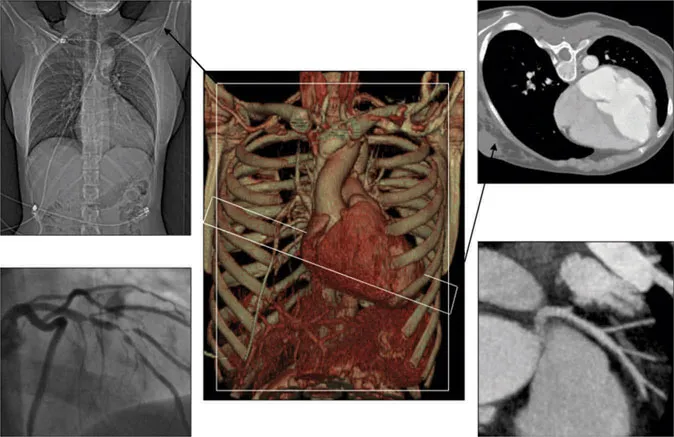
Figure 1.1 Planar versus tomographic imaging
The center panel of this figure shows a 3-D volume-rendered CT image of the chest. The panels on the left and right show planar and tomographic images, respectively. The standard chest X-ray (upper left panel) is a planar projection of the cardiac chambers. The intra-arterial injection of contrast material during coronary angiography selectively enhances the coronary arteries (lower left panel). However, similar to the chest X-ray, the angiogram is a planar image, projecting the silhouette of the contrast-filled coronary artery lumen. The tomographic CT images of the cardiac chambers (right upper panel) and of a coronary artery (right lower panel) allow the visualization of details not seen with planar imaging.
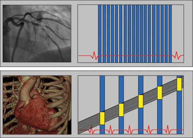
Figure 1.2 Image acquisition time, acquisition window
As demonstrated by the vertical bars in the upper part of the figure, cine-angiography acquires multiple image frames in one cardiac cycle. The time needed to acquire an individual planar image frame during cine-angiography is about 10 ms, allowing real-time imaging. In contrast, as shown in the lower part of the figure, the acquisition time for CT images is longer and is timed during late diastole of consecutive cardiac cycles. Minimal temporal resolution with multi-detector scanners is 135 ms (single source) and 75 ms (dual source).
Initial cardiovascular CT systems used electron-beam computed technology (EBCT). A rapidly oscillating X-ray beam was reflected onto a stationary tungsten target ring, encircling the patient. By eliminating the need to rotate the X-ray source mechanically around the patient, EBCT scanners were characterized by high temporal resolution.11 However, these scanners have been almost completely replaced by multi-detector systems (MDCT). In MDCT systems, the gantry (X-ray tube and detector) rotates rapidly around the patient. Initial single-detector CT systems, introduced in 1972 for body imaging, were limited by very slow rotation and long acquisition time. Fast gantry rotation, thin collimated detector rows, ECG-synchronized imaging, and acquisition of multiple slices per gantry rotation have since allowed the development of modern cardiovascular systems.12–14 Multi-slice scanners with 8, 16, 64, 256, and 320 slice acquisitions per rotation were introduced over the last decade.15–26 Dual-source scanners, with two X-ray tubes/detector systems, were introduced in 2008, and allowed a 50% reduction in temporal resolution.27,28 Today, high-end scanners permit rotation times as low as 270 ms, with resulting temporal resolution of 135 ms (single source) and 75 ms (dual source). Modern systems acquire data with a minimum collimated detector row width of 0.5 or 0.625 mm, resulting in isotropic spatial resolution of around 0.5 × 0.5 × 0.5 mm (Figure 1.3).
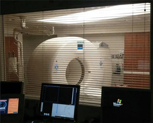
Figure 1.3 Multi-detector CT technology (MDCT)
This figure shows a second-generation dual-source scanner, photographed from the “control room.” Modern systems acquire up to 320 slices per rotation, with a minimum slice thickness below 0.75 mm, and spatial resolution about 0.5 mm.
The following chapters describe technical aspects of CT scan acquisition and evaluation, normal anatomy, and pathologic findings with MDCT in a variety of clinical conditions.
Chapter 2
Chapter CT perspective of normal cardiovascular anatomy
Because of the oblique orientation of the cardiovascular structures in the chest, cardiovascular imaging depends on reconstructions of defined image planes oblique to the body axes (z-axis), well known from echocardiography.29 With two-dimensional imaging modalities (e.g. standard echocardiography, most magnetic resonance sequences, and standard angiography), these image planes a...
Table of contents
- Cover
- Half Title
- Title Page
- Copyright Page
- Table of Contents
- Foreword
- Contributors
- Part 1 Basics of Multidetector Computed Tomography (MDCT)
- Part 2 Clinical Cardiovascular Applications
- References
- List of Videos
- Index
Frequently asked questions
Yes, you can cancel anytime from the Subscription tab in your account settings on the Perlego website. Your subscription will stay active until the end of your current billing period. Learn how to cancel your subscription
No, books cannot be downloaded as external files, such as PDFs, for use outside of Perlego. However, you can download books within the Perlego app for offline reading on mobile or tablet. Learn how to download books offline
We are an online textbook subscription service, where you can get access to an entire online library for less than the price of a single book per month. With over 1.5 million books across 990+ topics, we’ve got you covered! Learn about our mission
Look out for the read-aloud symbol on your next book to see if you can listen to it. The read-aloud tool reads text aloud for you, highlighting the text as it is being read. You can pause it, speed it up and slow it down. Learn more about Read Aloud
Yes! You can use the Perlego app on both iOS and Android devices to read anytime, anywhere — even offline. Perfect for commutes or when you’re on the go.
Please note we cannot support devices running on iOS 13 and Android 7 or earlier. Learn more about using the app
Please note we cannot support devices running on iOS 13 and Android 7 or earlier. Learn more about using the app
Yes, you can access Cardiac CT Made Easy by Paul Schoenhagen MD FAHA,Carl J. Schultz MD,Sandra S. Halliburton, Paul Schoenhagen MD FAHA, Carl J. Schultz MD, Sandra S. Halliburton in PDF and/or ePUB format, as well as other popular books in Medicine & Cardiology. We have over 1.5 million books available in our catalogue for you to explore.