- 81 pages
- English
- ePUB (mobile friendly)
- Available on iOS & Android
eBook - ePub
Ultrasound Assessment in Gynecologic Oncology
About this book
This innovative guide will help gynecologists or gynecologic surgeons to monitor the staging and progress of oncology treatment. An international expert here shows what office ultrasound can be used to achieve, how it correlates with other clinical findings, and how it can be integrated as necessary with other modalities and with the latest technological advances and developments in the field.
Trusted by 375,005 students
Access to over 1.5 million titles for a fair monthly price.
Study more efficiently using our study tools.
Information
Topic
Medicine1
Ultrasound Scanning of the Female Pelvis: Normal Findings
Introduction
Transvaginal ultrasound is currently considered as the first-line technique for imaging the female pelvis, especially for assessing the uterus and the adnexa. Transabdominal ultrasound is needed in some circumstances for assessing the entire pelvis or the abdomen for evaluating large structures or disease that spread intra-abdominally. Certainly, ultrasound has become an essential diagnostic tool for most clinicians involved in the clinical management of gynecological diseases, specifically tumoral entities.
In this chapter, we review the normal findings of the female pelvic organs as scanned by transvaginal ultrasound.
When assessing the female pelvis by ultrasound, we must clearly identify the anatomical landmarks to be assessed, especially when we focus on gynecological malignancies. From a practical perspective, the female pelvis can be divided into three parts: reproductive organs, nonreproductive organs, and pelvic wall structures.
Pelvic wall structures refer to the pelvic great vessels, muscles, and bones. Nonreproductive organs refer mainly to the bladder, ureter, recto-sigmoid, and bowel. Reproductive organs refer to the uterus, fallopian tubes, and ovaries. We should also include vaginal fornices, recto-vaginal septum, cardinal ligaments, or parametria and utero-sacral ligaments.
Pelvic Wall Structures
When assessing pelvic wall structures, we should image the great pelvic vessels, muscles, and bones. We focus on the pelvic vessels, as they are the main landmarks for pelvic scan in a gynecological oncological setting.
Pelvic vessels assessable by transvaginal ultrasound are mainly external iliac vessels (artery and vein), internal iliac vessels (artery and vein), and the uterine vessels (artery and venous plexus). Other vessels less frequently assessed by transvaginal ultrasound are the ovarian vessels. Due to the limitation of depth when using a high-frequency transvaginal probe, ovarian vessels are more difficult to assess. The identification of the great iliac vessels is essential for evaluating the presence of lymph nodes.
External iliac vessels are easily identified running parallel to the pelvic wall (Figure 1.1). The vein is larger than the artery and is located over the artery. The latter is clearly seen beating in virtually all women. It is important to insonate the vessels parallel to them in order to obtain a sagittal view of the vessels. This can be achieved by moving the endovaginal probe laterally and anteriorly.
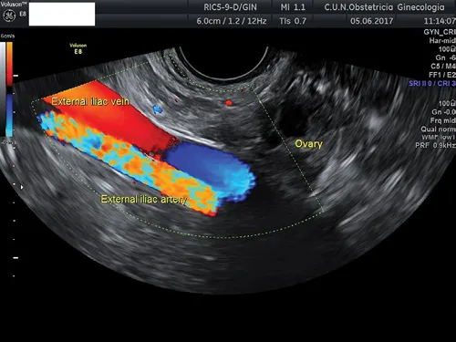
Figure 1.1 Transvaginal ultrasound showing right external iliac vessels. The ovary is seen lying over these vessels.
The internal iliac vessels are visible displacing the probe medially and posteriorly (Figure 1.2). Color Doppler may help in identifying these vessels.
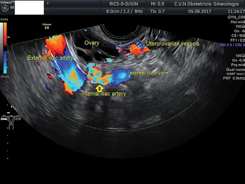
Figure 1.2 Transvaginal ultrasound depicting internal iliac vessels and utero-ovarian vessels.
Finally, the uterine vessels can be identified laterally to the cervix, either in the longitudinal or axial planes (Figure 1.3).
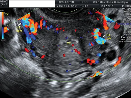
Figure 1.3 Transvaginal ultrasound showing both uterine arteries (UAs) at both sides of the cervix. The cervical canal (CC) can be observed.
Nonreproductive Organs
The bladder is easily identified as a central cystic structure located between the uterus and the abdominal wall (Figure 1.4). Bladder wall thickness can be measured, and the internal wall surface may be evaluated. It is a common finding to observe some irregularities of the bladder mucosa. When scanning an oncological patient, it is quite important to determine the presence of sliding of the bladder wall over the cervix, since this is a sign that indicates that the bladder wall is not involved, for example, in a case of cervical carcinoma.

Figure 1.4 Transvaginal ultrasound showing the uterus in the longitudinal plane. The bladder can be seen as an anechoic structure located anteriorly to the uterus.
The ureters can be seen passing through the bladder wall, and the ureteral meatus can be identified in both sides (Figure 1.5a). They are commonly identified as a hypoechoic creeping structure within the bladder wall (Figure 1.5b). More laterally, they can be observed crossing under the uterine artery (Figure 1.5c).
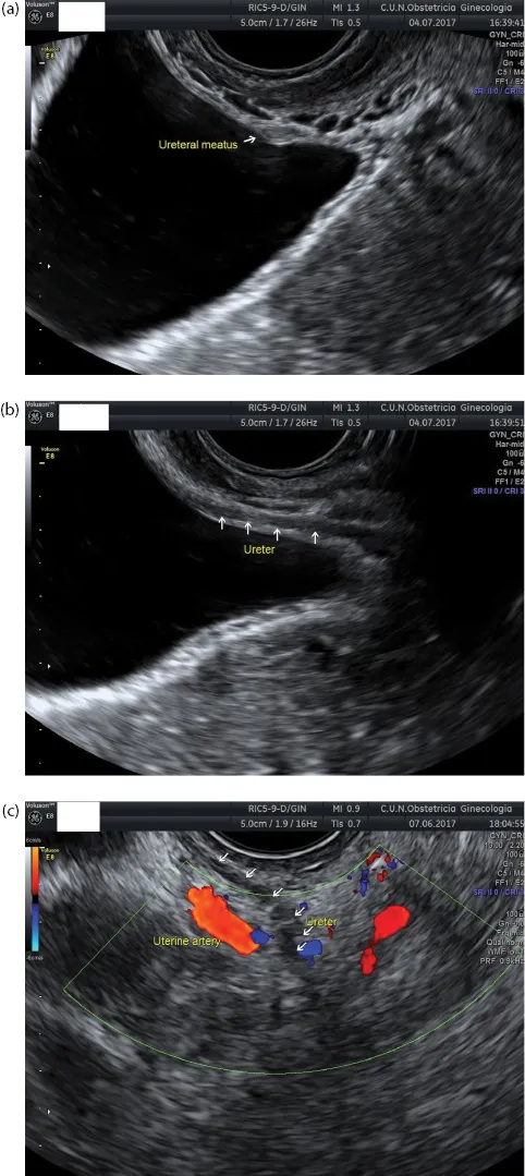
Figure 1.5 (a) Transvaginal ultrasound showing the bladder. The ureteral meatus can be observed in the longitudinal plane moving the endovaginal transducer laterally. (b) The transmural portion of the u...
Table of contents
- Cover
- Half Title
- Title Page
- Copyright Page
- Contents
- Foreword
- Preface
- 1. Ultrasound Scanning of the Female Pelvis: Normal Findings
- 2. Ultrasound for Differential Diagnosis of Adnexal Masses
- 3. Ultrasound Features of Ovarian Malignancies
- 4. Ultrasound Assessment of Intra-Abdominal Spread of Ovarian Cancer
- 5. Ultrasound Features of Endometrial Cancer
- 6. Ultrasound Features of Uterine Sarcomas
- 7. Ultrasound Assessment of Locoregional Spread of Endometrial Cancer
- 8. Ultrasound Features of Uterine Cervical Cancer
- 9. Ultrasound Features of Gestational Trophoblastic Disease
- 10. Ultrasound-Guided Procedures in Gynecologic Oncology
- 11. Ultrasound in Vulvar and Vaginal Cancer
- Index
Frequently asked questions
Yes, you can cancel anytime from the Subscription tab in your account settings on the Perlego website. Your subscription will stay active until the end of your current billing period. Learn how to cancel your subscription
No, books cannot be downloaded as external files, such as PDFs, for use outside of Perlego. However, you can download books within the Perlego app for offline reading on mobile or tablet. Learn how to download books offline
Perlego offers two plans: Essential and Complete
- Essential is ideal for learners and professionals who enjoy exploring a wide range of subjects. Access the Essential Library with 800,000+ trusted titles and best-sellers across business, personal growth, and the humanities. Includes unlimited reading time and Standard Read Aloud voice.
- Complete: Perfect for advanced learners and researchers needing full, unrestricted access. Unlock 1.5M+ books across hundreds of subjects, including academic and specialized titles. The Complete Plan also includes advanced features like Premium Read Aloud and Research Assistant.
We are an online textbook subscription service, where you can get access to an entire online library for less than the price of a single book per month. With over 1.5 million books across 990+ topics, we’ve got you covered! Learn about our mission
Look out for the read-aloud symbol on your next book to see if you can listen to it. The read-aloud tool reads text aloud for you, highlighting the text as it is being read. You can pause it, speed it up and slow it down. Learn more about Read Aloud
Yes! You can use the Perlego app on both iOS and Android devices to read anytime, anywhere — even offline. Perfect for commutes or when you’re on the go.
Please note we cannot support devices running on iOS 13 and Android 7 or earlier. Learn more about using the app
Please note we cannot support devices running on iOS 13 and Android 7 or earlier. Learn more about using the app
Yes, you can access Ultrasound Assessment in Gynecologic Oncology by Juan Luis Alcázar in PDF and/or ePUB format, as well as other popular books in Medicine & Gynecology, Obstetrics & Midwifery. We have over 1.5 million books available in our catalogue for you to explore.