Clinical Neuroscience offers a comprehensive overview of the biological bases of major psychological and psychiatric disorders, and provides foundational information regarding the anatomical and physiological principles of brain functioning. In addition, the book presents information concerning neuroplasticity, pharmacology, brain imaging, and brain stimulation techniques. Subsequent chapters address specific psychological disorders and neurodegenerative diseases, including major depressive and bipolar disorders, anxiety, schizophrenia, disorders of childhood origin, and addiction, as well as neurodegenerative disorders, such as Parkinson's and Alzheimer's diseases. This highly readable textbook expands case examples and illustrations to discuss the latest research findings in clinical neuroscience from an empirical, interdisciplinary perspective.
eBook - ePub
Clinical Neuroscience
Foundations of Psychological and Neurodegenerative Disorders
- 460 pages
- English
- ePUB (mobile friendly)
- Available on iOS & Android
eBook - ePub
About this book
Trusted by 375,005 students
Access to over 1.5 million titles for a fair monthly price.
Study more efficiently using our study tools.
Information
Subtopic
Mental Health in PsychologyIndex
Psychologyone
Neuroanatomy, Brain Development, Protection, Metabolic Needs of the Brain, and Neuroplasticity
Clinical neuroscience is a branch of neuroscience that focuses on the fundamental mechanisms that underlie disorders and diseases of the brain and central nervous system, and on scientifically based approaches to diagnosis and treatment. Clinical neuroscience has made its way into the mainstream media with daily headlines claiming, “Rewire your brain, overcome your addictions”, “train your mind, change your brain”, and online programs that offer personalized brain training programs to improve memory and cognition. This textbook will provide students with a foundation in neuroscience that will enable students to differentiate neuroscience fact from fiction and to navigate and decipher the clinical neuroscience literature. This chapter presents foundational information by reviewing the anatomy of the brain: the structures and associated functions of the forebrain, midbrain, and hindbrain, and lateralization of the hemispheres. It also covers glial cells and neurons, and addresses the processes involved in prenatal and postnatal brain development, including neurulation, dendritic arborization, and myelination. Sex differences in brain morphology are also explored. Finally, the concept of neuroplasticity is introduced with respect to the brain’s capacity to change in response to environmental stimulation, including cerebral stroke, developmental conditions, deprivation, limb amputation, and enrichment.
Chapter 1 Learning Objectives
- Identify the major divisions and subdivisions of the brain and nervous system.
- Describe the brain’s major structures and associated functions.
- Identify the major anatomical directional terms and planes of section.
- Distinguish between two main types of brain cells.
- Explain the process of brain development from prenatal to young adulthood.
- Distinguish between synaptogenesis, necrosis, and apoptosis.
- Define neuroplasticity and provide examples of plasticity.
- Explain physiological mechanisms of plasticity.
- Explain brain lateralization and provide specific examples of lateralization.
- Discuss brain morphology and sex differences.
- Identify protective layers of the brain.
- Discuss blood supply and metabolic needs of the brain.
Overview of the Nervous System
The nervous system has two main divisions: the peripheral nervous system (PNS) and the central nervous system (CNS) (Figure 1.1). The PNS is located outside the skull and spine, and detects environmental information via sensory receptors. It then transmits this information to the CNS by way of sensory nerves known as afferent nerves (from the Latin for carry information to the CNS). The PNS also transmits information from the CNS to muscles, glands, and internal organs by way of motor nerves known as efferent nerves (from the Latin for carry information away from the CNS) (Figure 1.2). The PNS is subdivided into the somatic and autonomic nervous systems. The somatic division includes both sensory and motor nerves, and controls skeletal and voluntary movement. The autonomic division controls glands and muscles of internal organs, and regulates internal bodily processes. The autonomic division consists of three main parts: the sympathetic (arousal, “fight or flight”) and parasympathetic (restoration) nervous systems, as well as the enteric nervous system (ENS) that governs the function of the gastrointestinal system. The ENS receives sympathetic and parasympathetic input but is also capable of acting independently to affect the functions of the gastrointestinal tract. The gastrointestinal tract has recently been implicated in depression symptoms. Major depressive disorder will be covered in detail in Chapter 6.
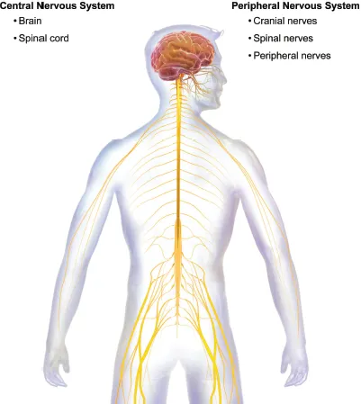
Figure 1.1. Organization of the Nervous System
Copyright Blausen Medical Communications. Reproduced by permission.
Figure 1.2. Afferent and Efferent Nerves
Reproduced by permission.
BOX 1.1 Can Probiotics Help Lessen Depression Symptoms?
The human gastrointestinal (GI) tract consists of a large number of microorganisms. Research has found a bidirectional relationship between the brain and the GI tract that involves neural, hormonal, and immunological pathways. Preliminary evidence suggests that these microbes may play an important role in the development and maturation of brain systems that are associated with stress responses. Recently, Slykerman et al. (2017) found that women who were treated with a probiotic for six months following childbirth reported significantly less depression and anxiety symptoms relative to women who were randomly assigned to a placebo condition. In patients with irritable bowel syndrome (IBS), Pinto-Sanchez and colleagues (2017) found probiotic treatment was associated with a significant reduction in depression and increased quality of life in patients with IBS compared to patients taking a placebo. Furthermore, the clinical improvements were associated with changes in brain activation patterns—i.e., reduced limbic reactivity to negative emotional stimuli. Although additional studies are needed, researchers hypothesize that probiotics lead to a reduction in GI inflammation while simultaneously increasing brain levels of serotonin.
The CNS is located in the skull and spine, and consists of the brain and the spinal cord. Twelve pairs of cranial nerves and 31 pairs of spinal nerves connect the PNS to the brain and spinal cord. As shown in Figure 1.3, cranial nerves are located on the ventral surface of the brain and involve numerous functions of the head, neck, and face. Cranial nerves I and II (olfactory and optic) are located in the forebrain, while cranial nerves III and IV (oculomotor, trochlear) are located in the midbrain. The final eight pairs of cranial nerves, which are found on the ventral surface of the brain stem, are important in tongue and neck movements, as well as regulating internal organs and vital functions.
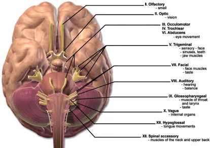
Figure 1.3. Location and Function of Cranial Nerves
Copyright Blausen Medical Communications. Reproduced by permission.
Brain Regions, Structures, and Functions
Directionality and Terminology
The brain can be viewed from various anatomical directions based on three axes: anterior-posterior, dorsal-ventral, and medial-lateral. Anterior refers to the front (also known as rostral in four-legged animals) and posterior refers to the rear or tail (also known as caudal). Dorsal refers to the back or top and ventral toward the belly or ground. Lateral refers to the side and medial the midline (Figure 1.4). Ipsilateral refers to the same side of the body and contralateral to the opposite side of the body. The structures of the brain can also be viewed from several sections (“cuts”): horizontal, sagittal, and coronal planes. A horizontal plane runs parallel to the top of the brain, a sagittal plane runs parallel to the midline of the brain, and a coronal plane runs parallel to the front of the brain, dividing the nervous system from front to back (“frontal cut”). A section that divides the brain into two equal halves is known as a midsagittal section. This directional terminology will be important for understanding the information to follow (Tables 1.1 and 1.2) as well as understanding and interpreting the scientific literature.
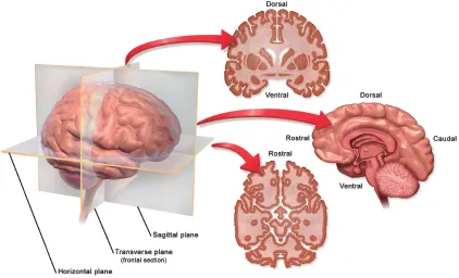
Figure 1.4. Directionality and Planes
Copyright Blausen Medical Communications. Reproduced by permission.
| Cerebrum | The cerebrum is the largest part of the human brain. It is associated with higher brain functions and is divided into four lobes: frontal, parietal, occipital, and temporal. |
| Frontal Lobe | The front lobes are involved in executive functions, planning, speech production, motor movements, emotional regulation, and complex problem solving. |
| Parietal Lobe | The parietal lobes are involved in somatosensation, spatial orientation, and sensory integration. |
| Occipital Lobe | The occipital lobes are involved in processing and perception of visual stimuli. |
| Temporal Lobe | The temporal lobes are involved in perception and processing of auditory stimuli, memory, speech pattern recognition, and receptive language. |
| Cerebellum | The cerebellum, like the cerebrum, has two hemispheres. The cerebellum is involved in numerous functions, including memory, coordination of movement, posture, and balance. |
| Limbic System | The limbic system is a set of interconnected structures involved in emotions, learning, motivation, memory, and the four Fs. |
| Thalamus | The thalamus functions as the major relay station of the brain for sensory-motor information and is involved in sleep and arousal. |
| Hypothalamus | The hypothalamus is involved in hormonal regulation and numerous homeostatic functions, such as temperature regulation, circadian rhythms, thirst, and hunger. |
| Amygdala | The amygdala is involved in memory, processing and formations of emotion, and motivation. |
| Hippocampus | The hippocampus is involved in memory formation and memory retrieval, as well as spatial navigation. |
| Brain Stem | The brain stem connects the cerebrum to the spinal cord, has numerous ascending and descending sensory-motor pathways, and is involved in maintaining vital functions. |
| Midbrain/Mesencephalon | The midbrain is the most rostral part of the brain stem. It includes the tectum and tegmentum, and is involved in vision, hearing, eye movement, and voluntary body movement. |
| Pons | The pons is part of the hindbrain and is involved in motor control, posture, and sensory-motor functions. |
| Medulla | The medulla is in the most caudal part of the brain stem located between the pons and spinal cord, and is involved in regulating autonomic functions, such as breathing, heart rate, and blood pressure. |
Brain Divisions
The brain consists of three major divisions with further subdivisions: 1) prosencephalon, telencephalon, and diencephalon; 2) mesencephalon; and 3) rhombencephalon, metencephalon, and myelencephalon. The prosencephalon refers to the forebrain, mesencephalon the midbrain, and the rhombencephalon the hindbrain (Figure 1.5).
Table 1.2Major Divisions of the Brain: Structures and Ventricles
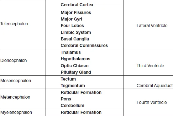
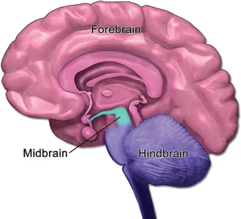
Figure 1.5. Sagittal Section Depicting Three Primary Divisions of the Brain
Copyright Blausen Medical Communications. Reproduced by permission.
Forebrain and Lateralization
The prosencephalon—i.e., the forebrain—consists of the telencephalon and diencephalon. The telencephalon is the largest division of the human brain and is involved in developing complex cognitive and behavioral processes, such as initiating movement, interpreting sensory stimulation, and higher-level cognition such as planning and problem solving (i.e., executive functions), and language. The telencephalon is made up of two cerebral hemispheres—the right and the left (Figure 1.6)—which are connected by a number of bundles of nerve fibers (i.e., commissures), including the corpus callosum, anterior, posterior, hippocampal, and habenular commissures. The commissures act as a conduit through which the right and left hemispheres exchange information and function interdependently (Springer & Deutsch, 1993). The hemispheres are asymmetrical and vary in size depending on specific structures or regions (e.g., left frontal is larger in size than right frontal; Watkins et al., 2001). The hemispheres traditionally have been described as specialized in function (lateralization), and functions traditionally ascribed to the left hemisphere include language-related functions, logical thinking, and writing; visuospatial, musical, and artistic abilities have been ascribed to the right hemisphere (Springer & Deutsch, 1993). Early evidence of these processing hemispheric differences derived largely from individuals with brain damage, surgically cut corpus callosum (“split-brain patients”), and neuropsychological studies. For example, damage to the left frontal lobe (Broca’s area) or left temporal lobe (Wernicke’s area) can result in difficulties with language production and comprehension (Damasio, 1991); however, this is not always the case and variability exists among patients. Damage to the right hemisphere can result in spatial reasoning difficulties, such as judging line orientation (Benton, Hannay, & Varney, 1975) and interpreting facial expressions (D. Bowers et al., 1985). Patients with uncontrollable forms of epilepsy who have had their corpus callosum surgically severed have been studied extensively by Michael Gazzaniga of UC Santa Barbara. Gazzaniga’s work has further substantiated lateralization of cognitive functions (e.g., Gazzaniga, 2005). Many other deficits have been attributed to left and right hemisphere damage and vary depending on the cortical and subcortical regions involved, as well as factors such as age, sex, intelligence, location, and severity of the damage (Kolb, 1989; Kolb & Whishaw, 1996).
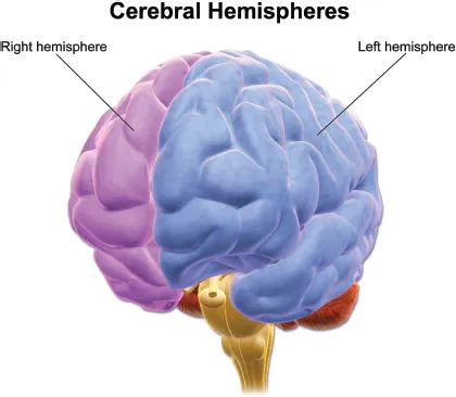
Figure 1.6. Left and Right Hemispheres of the Brain
Copyright Blausen Medical Communications. Reproduced by permission.
More recently, brain imaging technology has been used to explore lateralized functions in healthy individuals across a spectrum of cognitive processes. For example, studies have found that in most people, the left hemisphere is primarily activated when viewing static emotional facial expressions, particularly positive emotions, while the right hemisphere is correlated with negative emotions and dynamic facial expressions (Baeken et al., 2010). Spatial movements, particularly global movements are largely right hemisphere dominant (Floegel & Kell, 2017). Language and reading are largely, although not exclusively, left hemisphere dominant in both males and females, particularly in right-handed individuals (Nenert et al., 2017; Waldie et al., 2017). Interestingly, in congenitally blind individuals, a reduction is found in left hemisphere lateralization when performing language-related tasks (Lane et al., 2017). These findings support that the neurobiology of language can be modified by experience and are consistent with the previous discussion of neuroplasticity. Additional cognitive processes (e.g., musical ability, memory) have been investigated with respect to lateralization, and the current view is that constructs such as language, memory, and emotion consist of an array of individual cognitive processes that often require involvement of both hemispheres. The relative contribution of each hemisphere involved in various cognitive processes remains under investigation, however, as well as factors that may mitigate hemispheric involvement. In terms of hemispheric control of the body, the left hemisphere generally controls the right side of the body and the right hemisphere controls the left side of the body (i.e., contralateral control).
The hemispheres are covered by the cerebral cortex and separated by the longitudinal fissure (Figure 1.7). The cerebral cortex is approximately 3 mm thick, convoluted, and consists of six layers of cells (of varying thickness) running parallel to its surface (Martini, 1998; List et al., 2013; Tamnes et al., 2017). Because the cortex consists mainly of cell bodies, it has a grayish appearance and is commonly referred to as “gray matter”. Extensions of the cell body of neurons (axons) project to other areas of the cortex and to subcortical regions of the brain, and are often covered in myelin. Myelin consists of fats and proteins, has a whitish appearance, and is commonly referred to as “white matter”. According to Martini (1998), the total surface area of the cortex is roughly equivalent to 2.5 square feet of flat surface. The size and shape of the skull cause the cortical structure of the brain to fold inward. Hence much of the brain is hidden within the grooves. These folds or bumps are called gyri, and the grooves are known as sulci (smaller grooves) and fissures (major grooves). The largest fissure is the longitudinal fissure that separates the left and right hemispheres. The lat...
Table of contents
- Cover
- Half Title
- Title
- Copyright
- Dedication
- Contents
- Acknowledgments
- Foreword
- Preface
- Chapter 1 — Neuroanatomy, Brain Development, Protection, Metabolic Needs of the Brain, and Neuroplasticity
- Chapter 2 — Cellular Function, Neurotransmission, and Pharmacology
- Chapter 3 — Techniques of Brain Imaging and Brain Stimulation
- Chapter 4 — Neurocognitive Disorder Due to Dementia of Alzheimer’s Type and Parkinson’s Diseases
- Chapter 5 — Schizophrenia
- Chapter 6 — Major Depressive Disorder and Bipolar Disorder
- Chapter 7 — Anxiety Disorder and Obsessive-Compulsive Disorder
- Chapter 8 — Addiction and Substance Use Disorders
- Chapter 9 — Disorders of Childhood Origin: Attention Deficit Hyperactivity Disorder, ASD, and Tourette’s Disorder
- References
- Index
Frequently asked questions
Yes, you can cancel anytime from the Subscription tab in your account settings on the Perlego website. Your subscription will stay active until the end of your current billing period. Learn how to cancel your subscription
No, books cannot be downloaded as external files, such as PDFs, for use outside of Perlego. However, you can download books within the Perlego app for offline reading on mobile or tablet. Learn how to download books offline
Perlego offers two plans: Essential and Complete
- Essential is ideal for learners and professionals who enjoy exploring a wide range of subjects. Access the Essential Library with 800,000+ trusted titles and best-sellers across business, personal growth, and the humanities. Includes unlimited reading time and Standard Read Aloud voice.
- Complete: Perfect for advanced learners and researchers needing full, unrestricted access. Unlock 1.5M+ books across hundreds of subjects, including academic and specialized titles. The Complete Plan also includes advanced features like Premium Read Aloud and Research Assistant.
We are an online textbook subscription service, where you can get access to an entire online library for less than the price of a single book per month. With over 1.5 million books across 990+ topics, we’ve got you covered! Learn about our mission
Look out for the read-aloud symbol on your next book to see if you can listen to it. The read-aloud tool reads text aloud for you, highlighting the text as it is being read. You can pause it, speed it up and slow it down. Learn more about Read Aloud
Yes! You can use the Perlego app on both iOS and Android devices to read anytime, anywhere — even offline. Perfect for commutes or when you’re on the go.
Please note we cannot support devices running on iOS 13 and Android 7 or earlier. Learn more about using the app
Please note we cannot support devices running on iOS 13 and Android 7 or earlier. Learn more about using the app
Yes, you can access Clinical Neuroscience by Lisa Weyandt in PDF and/or ePUB format, as well as other popular books in Psychology & Mental Health in Psychology. We have over 1.5 million books available in our catalogue for you to explore.