Point of Care Ultrasound Made Easy (POCUSME) is an exciting and innovative book that aims to teach all healthcare professionals how to do simple and clinically relevant ultrasound scanning at the point of care. This book will help you solve clinical problems at the bedside across a range of specialty areas, including: trauma, emergency medicine, respiratory medicine, cardiology, general surgery, otolaryngology and vascular surgery. Straightforward and practical, and designed for clinicians who are generally unfamiliar with ultrasound scanning, it will make a positive difference to your clinical practice, and help improve the delivery of optimised patient care. So read the book, grab an ultrasound machine, and please embrace the Point of Care Ultrasound movement!
eBook - ePub
Point of Care Ultrasound Made Easy
- 154 pages
- English
- ePUB (mobile friendly)
- Available on iOS & Android
eBook - ePub
Point of Care Ultrasound Made Easy
About this book
Trusted by 375,005 students
Access to over 1.5 million titles for a fair monthly price.
Study more efficiently using our study tools.
Information
Part I
Background to Point-of-Care Ultrasound Made Easy (POCUSME)
Chapter 1
How Does Ultrasound Work?
John McCafferty
Learning Objectives
What Is Ultrasound?
Properties of Ultrasound in Tissues
Ultrasound Imaging
Image Acquisition
Scanning Modes
Optimising Imaging
Other Functions
Ultrasound Devices Used in Point of Care
Ultrasound Safety
Learning Objectives
◾Understand the basic physics behind medical ultrasound imaging.
◾Understand the hardware components of a medical ultrasound scanner.
◾Understand the different imaging modes: B-mode, M-mode and Doppler.
◾Know how to acquire an ultrasound image, appreciating the imaging conventions.
◾Know how to optimise your ultrasound image.
◾Appreciate your choices of point-of-care ultrasound (POCUS) device.
◾Have an awareness of safety factors in POCUS.
What Is Ultrasound?
Ultrasound waves are mechanical energy that are transmitted through a medium from repetitive periodic oscillations of a transducer. The number of such cycles per second is termed the frequency of the ultrasound signal. Ultrasound refers to sound propagated at frequencies higher than the audible spectrum for humans (>20 KHz) (see Figure 1.1). Typically, medical ultrasound imaging operates in the range 3.5–20 MHz.
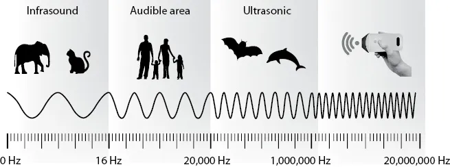
Figure 1.1 The frequency spectrum of sound waves with reference to the medical ultrasound range.
Properties of Ultrasound in Tissues
A B-mode ultrasound image is constructed from echoes generated by reflection of ultrasound from boundaries and scattering from small inhomogeneities within the tissue structures. Reflection occurs at the boundaries between two mediums of different acoustic impedance such as those between organs. Here, acoustic impedance is defined as the product of density of the tissue and the speed of sound within the tissue. The amplitude of the ultrasound wave that is reflected back into the tissue/organ or transmitted across a tissue/organ boundary is dependent on the change in acoustic impedance. If the change is small, the majority of the ultrasound will be transmitted and little reflected; if the change is large, the majority will be reflected back and little transmitted. For example, the medium of fluid or water has low impedance and will conduct the ultrasound wave, whereas bone is a poor conductor and will reflect the ultrasound wave back towards its source (see Figure 1.2).
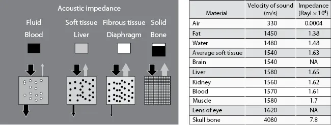
Figure 1.2 Impedance values of typical tissues seen in the body. Air has very low impedance, meaning the ultrasound wave is strongly reflected at an air/tissue interface. This is why ultrasound gel is used when scanning a patient to avoid air interfaces between the transducer and the patient’s skin.
Small inhomogeneities within tissues/organs result in scattering of the ultrasound beam. This scattering is dependent on the size of the scatterer relative to the frequency/wavelength of the ultrasound beam. However, the scattering from these small structures (including red blood cells) is much smaller than typical reflections from boundaries so that within a B-mode image, boundaries are associated with brighter echoes compared to the less bright echoes generated by scattering within soft tissue.
Ultrasound Imaging
Ultrasound imaging uses the differences in the acoustic properties of tissues to build a picture of the structures within the body. The ultrasound signal is generated by a transducer that contains an array of tiny piezoelectric crystals which when excited by an electrical signal vibrate at a set frequency and transmit an ultrasound signal. The transducer is also a receiver, detecting the re...
Table of contents
- Cover
- Half Title
- Title Page
- Copyright Page
- Dedication
- Contents
- Foreword
- Acknowledgements
- Contributors
- Part I: Background to Point-of-Care Ultrasound Made Easy (POCUSME)
- Part II: Ultrasound Assessments by Body System
- Part III: Assessment
- Index
Frequently asked questions
Yes, you can cancel anytime from the Subscription tab in your account settings on the Perlego website. Your subscription will stay active until the end of your current billing period. Learn how to cancel your subscription
No, books cannot be downloaded as external files, such as PDFs, for use outside of Perlego. However, you can download books within the Perlego app for offline reading on mobile or tablet. Learn how to download books offline
Perlego offers two plans: Essential and Complete
- Essential is ideal for learners and professionals who enjoy exploring a wide range of subjects. Access the Essential Library with 800,000+ trusted titles and best-sellers across business, personal growth, and the humanities. Includes unlimited reading time and Standard Read Aloud voice.
- Complete: Perfect for advanced learners and researchers needing full, unrestricted access. Unlock 1.5M+ books across hundreds of subjects, including academic and specialized titles. The Complete Plan also includes advanced features like Premium Read Aloud and Research Assistant.
We are an online textbook subscription service, where you can get access to an entire online library for less than the price of a single book per month. With over 1.5 million books across 990+ topics, we’ve got you covered! Learn about our mission
Look out for the read-aloud symbol on your next book to see if you can listen to it. The read-aloud tool reads text aloud for you, highlighting the text as it is being read. You can pause it, speed it up and slow it down. Learn more about Read Aloud
Yes! You can use the Perlego app on both iOS and Android devices to read anytime, anywhere — even offline. Perfect for commutes or when you’re on the go.
Please note we cannot support devices running on iOS 13 and Android 7 or earlier. Learn more about using the app
Please note we cannot support devices running on iOS 13 and Android 7 or earlier. Learn more about using the app
Yes, you can access Point of Care Ultrasound Made Easy by John McCafferty, James M Forsyth, John McCafferty,James Forsyth,James M Forsyth in PDF and/or ePUB format, as well as other popular books in Medicine & Clinical Medicine. We have over 1.5 million books available in our catalogue for you to explore.