eBook - ePub
Infectious Diseases of the Dog and Cat
A Color Handbook
- 320 pages
- English
- ePUB (mobile friendly)
- Available on iOS & Android
eBook - ePub
Infectious Diseases of the Dog and Cat
A Color Handbook
About this book
This is a concise and quick reference guide, clinically oriented, based on experience underpinned by published research data. The systems-based approach emphasizes the problem list process utilized by most practitioners when faced with a patient presenting with a single or variety of clinical symptoms. This allows generation of a complete list of infectious disease differentials, and the ability to include or exclude those indicated based on patient risk assessment. Pathogen specific information enables rational choice of diagnostics, therapy, and prognostication for a complete list of small animal infectious disease that includes bacterial, viral, parasitic/protozoal and fungal.
Trusted by 375,005 students
Access to over 1.5 million titles for a fair monthly price.
Study more efficiently using our study tools.
Information
Topic
MedicineSubtopic
Diseases & AllergiesCHAPTER 1
RESPIRATORY DISEASES
AELUROSTRONGYLUS ABSTRUSUS/CAT LUNGWORM, VERMINOUS PNEUMONIA
Dennis Spann
Definition
The metastrongyloid nematode Aelurostrongylus abstrusus causes lower airway disease in cats.1 Infected cats cough secondary to inflammation from the parasite lodging in the terminal bronchioles and alveolar ducts.
Etiology and pathogenesis
Aelurostrongylus infestation occurs when a cat (definitive host) or dog consumes an intermediate (snail or slug) or paratenic host (frogs, lizards, birds, and rodents) with encysted larvae. The larvae travel through the lymphatics before entering the lung parenchyma and bronchioles. They penetrate the lung tissue deeply but extrude eggs into the bronchioles and alveolar ducts. The eggs then hatch into larvae, which travel up the mucociliary escalator and are swallowed; they exit via the gastrointestinal tract and pass into the environment (Fig. 1.1). Intermediate hosts, snails and slugs (Fig. 1.2), are penetrated by the L1 larvae, which then develop into L3 larvae. The intermediate host may be consumed by a paratenic host, in which the larvae encyst. Infection of the definitive host can occur via ingestion of the intermediate or paratenic host.1, 2
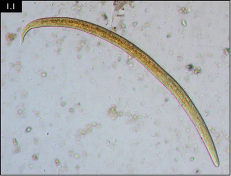
Fig. 1.1 L1 Aelurostrongylus abstrusus larvae from fecal float. (Courtesy of Drs Donato Traversa and Angela Di Cesare)

Fig. 1.2 Cornu aspersum, the garden snail, is a common intermediate host for A. abstrusus. (Courtesy of Rasbak, https://commons.wikimedia.org/wiki/File:Cornu_aspersum_(Segrijnslak).webp)
Geography
The parasite occurs worldwide.
Incidence and risk factors
The prevalence of larvae in feces has been estimated at 1.2% in indoor owned cats and up to 50% in free roaming cats in some areas.1–3 Risk factors include hunting and increased time outdoors. Older cats may have higher prevalence due to cumulative hunting time.
Clinical signs
Cats may be clinically normal if there is a low parasite burden. Disease is characterized by varying degrees of coughing, wheezing, respiratory distress, open-mouth breathing, abdominal effort and mucopurulent nasal discharge.3 Debility and even death can occur in rare cases with severe infections or comorbidities.3
Diagnosis
Diagnosis is most often based on detection of larvae in feces via the Baermann technique. The larvae are approximately 360–400 μm in length and the tail ends in a kink with a dorsal subterminal spine (Fig. 1.1). Fecal floatation is less sensitive than Baermann testing, with zinc sulfate flotation having a higher yield than sucrose flotation. Thoracic radiographs are usually unremarkable, but a nodulointerstitial pattern can be present in severe cases (Fig. 1.3).
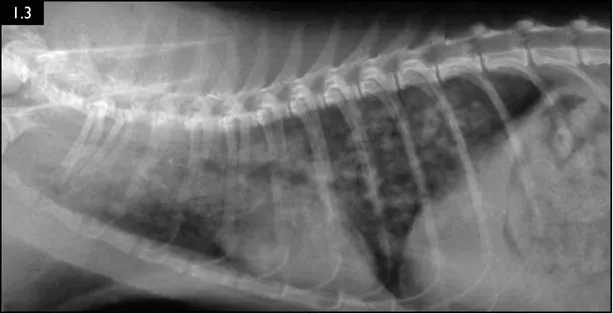
Fig. 1.3 Radiograph of a feline patient with a severe A. abstrusus infection. (Courtesy of Dr Maria Grazia Pennisi)
Therapy
Fenbendazole (50 mg/kg/day, PO, for 10–14 days), ivermectin (400 µg/kg, SC, twice at a 3-week interval) or an emodepside 2.1%/praziquantel 8.6% spot-on formulation can be used.3, 4 Anti-inflammatory doses of a corticosteroid can be used in severe infections.
Prognosis/complications
Prognosis is fair with treatment. In rare instances, heavy infestations may cause tissue damage, pneumothorax or hydrothorax.
Prevention
Prevention of predation (hunting) is the main control measure.
BACTERIAL PNEUMONIA
J Scott Weese
Definition
Bacterial pneumonia is an infectious process occurring within lung parenchyma.
Etiology and pathogenesis
Disease occurs secondary to events such as progression of upper respiratory tract infection, aspiration, foreign body migration and hematogenous spread. The end result is a bacterial load within the lung parenchyma and airways beyond a level that inherent protective mechanisms and the immune system can manage. Factors such as the degree of bacterial burden, presence of debris (e.g. aspirated material), compromise of local immune defenses and systemic immunocompromise influence whether disease occurs, and its severity.
A range of bacteria can be involved, most commonly Enterobacteriaceae (e.g. Escherichia coli, Enterobacter spp., Klebsiella spp.), Pasteurella spp., Bordetella bronchiseptica, Streptococcus spp. and anaerobes.5 Mycoplasma spp. are commonly identified but their role in disease is unclear, particularly in dogs.
Incidence and risk factors
The incidence is poorly described. It is a relatively common problem in dogs with megaesophagus, diseases that compromise laryngeal or pharyngeal function, or underlying pulmonary diseases (e.g. ciliary dyskinesia, bronchiectasis). Secondary pneumonia is uncommon following viral or bacterial upper respiratory tract infection but will develop in a small percentage of cases.
Clinical signs
It is important to differentiate upper respiratory tract infection from pneumonia. Dogs and cats with pneumonia typically have more severe signs. These indicate systemic disease and disease of the pulmonary parenchyma, and include fever, lethargy, decreased appetite, increased respiratory rate and effort, and auscultable crackles and wheezes. Signs of systemic inflammation may also be present.
Diagnosis
Clinical signs can be strongly suggestive of bacterial pneumonia, but radiographs are important for confirmation, to characterize the disease (and potential etiology) and to provide a baseline for monitoring response to treatment (Figs. 1.4–1.6). A lag between clinical signs and radiographic changes can occur, and initial radiographs may be normal or appear discordant with clinical severity.
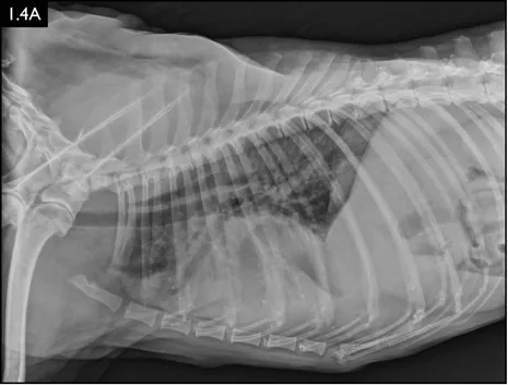
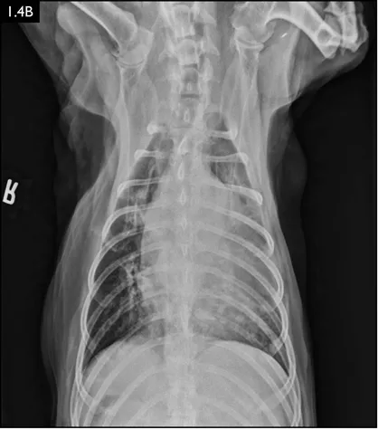
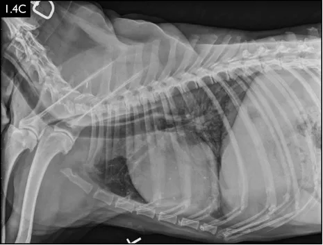
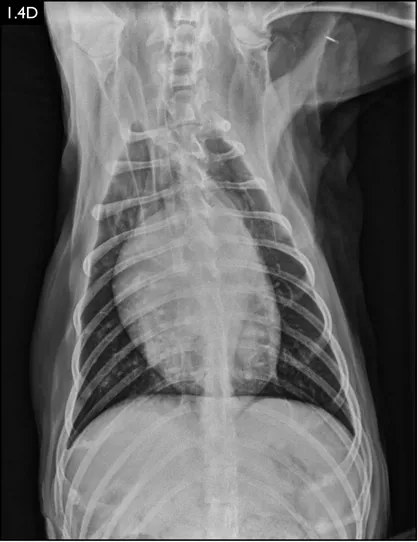
Fig. 1.4A–D Lateral and ventrodorsal radiographs of a dog with doxycycline-responsive pneumonia of unknown etiology before treatment ( A, B) and six days later ( C, D). Note the severe multilobar alveolar pattern that was present initially, most prominently in the left cranial lung lobe. (Courtesy of Atlantic Veterinary College)
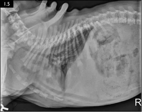
Fig. 1.5 Radiograph showing severe tracheal narrowing and concurrent bronchoalveolar pneumonia in a bulldog puppy with severe respiratory compromise. (Courtesy of Atlantic Veterinary College)
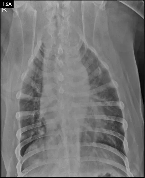
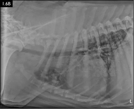
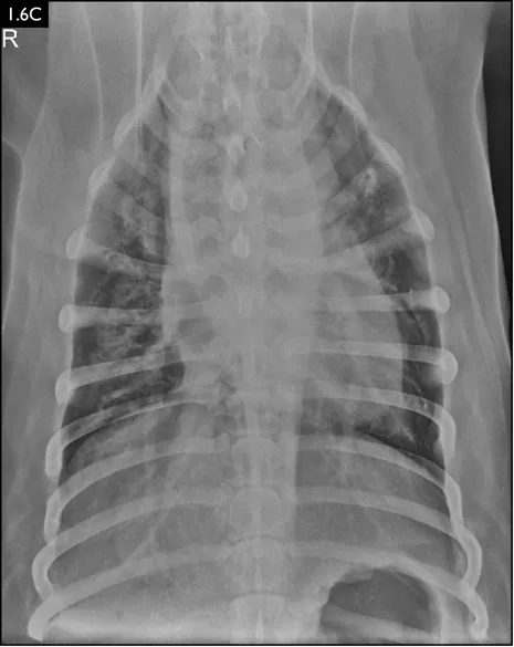
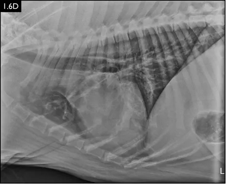
Fig. 1.6A–D Radiographs of a dog with suspected aspiration pneumonia. A diffuse alveolar pulm...
Table of contents
- Cover
- Title Page
- Copyright Page
- Dedication Page
- Contents
- Preface
- Contributors
- Abbreviations
- CHAPTER 1 RESPIRATORY DISEASES
- CHAPTER 2 GASTROINTESTINAL DISEASES
- CHAPTER 3 NEUROLOGIC DISEASES
- CHAPTER 4 GENITOURINARY DISEASES
- CHAPTER 5 SKIN AND SOFT TISSUE DISEASES
- CHAPTER 6 MULTISYSTEM DISEASES
- Index
Frequently asked questions
Yes, you can cancel anytime from the Subscription tab in your account settings on the Perlego website. Your subscription will stay active until the end of your current billing period. Learn how to cancel your subscription
No, books cannot be downloaded as external files, such as PDFs, for use outside of Perlego. However, you can download books within the Perlego app for offline reading on mobile or tablet. Learn how to download books offline
Perlego offers two plans: Essential and Complete
- Essential is ideal for learners and professionals who enjoy exploring a wide range of subjects. Access the Essential Library with 800,000+ trusted titles and best-sellers across business, personal growth, and the humanities. Includes unlimited reading time and Standard Read Aloud voice.
- Complete: Perfect for advanced learners and researchers needing full, unrestricted access. Unlock 1.5M+ books across hundreds of subjects, including academic and specialized titles. The Complete Plan also includes advanced features like Premium Read Aloud and Research Assistant.
We are an online textbook subscription service, where you can get access to an entire online library for less than the price of a single book per month. With over 1.5 million books across 990+ topics, we’ve got you covered! Learn about our mission
Look out for the read-aloud symbol on your next book to see if you can listen to it. The read-aloud tool reads text aloud for you, highlighting the text as it is being read. You can pause it, speed it up and slow it down. Learn more about Read Aloud
Yes! You can use the Perlego app on both iOS and Android devices to read anytime, anywhere — even offline. Perfect for commutes or when you’re on the go.
Please note we cannot support devices running on iOS 13 and Android 7 or earlier. Learn more about using the app
Please note we cannot support devices running on iOS 13 and Android 7 or earlier. Learn more about using the app
Yes, you can access Infectious Diseases of the Dog and Cat by Scott Weese, Michelle Evason, Scott Weese,Michelle Evason,J Scott Weese in PDF and/or ePUB format, as well as other popular books in Medicine & Diseases & Allergies. We have over 1.5 million books available in our catalogue for you to explore.