- 206 pages
- English
- ePUB (mobile friendly)
- Available on iOS & Android
eBook - ePub
About this book
This highly illustrated book brings together many concepts related to skin care and antioxidant usage in one convenient text. The second edition now contains the latest antioxidants being marketed, and an analysis of risks and benefits associated.
Trusted by 375,005 students
Access to over 1.5 million titles for a fair monthly price.
Study more efficiently using our study tools.
Information
Structure and function of skin | 1 |
Skin is key to our biology, our sensory experiences, our information gathering, and our relationships with others.
—Nina G. Jablonski
Skin: A Natural History
As the largest organ of the body, the skin provides a protective barrier for the body against the external world and also prevents transepidermal water loss (TEWL), permitting an aqueous organism to live in a rather arid environment. In addition, there are many other functions provided by skin that are often overlooked. For example, the skin maintains body temperature through two mechanisms: one in which the body is cooled by the action of sweating, via sweat glands; the other by the vasoconstriction or vasodilation of the blood vessels in the dermis, which either decreases or increases the flow of blood to the dermis from other internal organs. If the internal organs are overheated, vasodilation occurs in the dermis, allowing increased blood flow, which in turn releases heat to the environment. Through the combined process of sweating, sebum production, and desquamation, a thin film is present on the surface skin, protecting it from bacterial infections and invasion by foreign substances, as well as hydrating and lubricating the surface. Another essential function of skin is sensation, which is provided by a complex network of nerves located in the dermis and at the base of the epidermis.
Morphologically, the skin is composed of two principal components, the epidermis and dermis, which contain various cell types and structural proteins. The hypodermis—also known as the subcutaneous layer—is often categorized as a third component, located below the dermis. Primarily it is composed of adipose tissue. The epidermis is a squamous epithelium that contains several appendages: pilosebaceous unit, nail, and sweat gland. The dermis provides the skin with mechanical strength and elasticity, which is brought about by collagen and elastin. A diagram of skin is provided in Figure 1.1, where the structural components of the dermis and epidermis are shown as well as the various appendages of skin and its vascular network.
In the remainder of this chapter, we review the intricate structural components of the epidermis and dermis as well as the dermal-epidermal junction—the structure that connects the two skin layers. In addition, we discuss the morphology of various appendages of skin, such as the pilosebaceous unit (including both the hair follicle and sebaceous gland), nail unit, eccrine glands, and apocrine glands. Since skin plays such an integral role in immune function, we will survey some fundamental elements of the immune system as it involves skin. Many of the topics touched upon in this chapter are important for our later discussions on free radical mechanisms and antioxidants in skin.
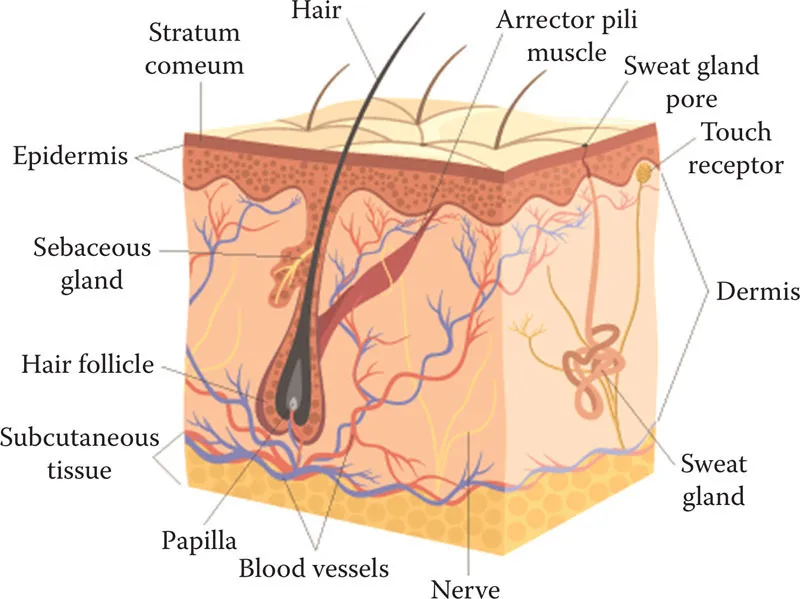
Figure 1.1 Structure of human skin.
EPIDERMIS
This layer of the skin primarily consists of keratinocyte cells, which undergo a process of events leading to terminal differentiation. The keratinocyte begins its journey as a metabolically active cell at the base of the epidermis and eventually makes it way to the outermost layer of skin, where it becomes void of its normal cellular organelles, becomes filled with keratin intermediate filaments (KIFs) and matrix proteins, and contains an unusually tough, protein-rich plasma membrane—known as the cell envelope—which is highly cross-linked. Through this process, the keratinocyte drastically changes shape during its voyage from the bottom to the top of the epidermis. Meanwhile, new organelles are synthesized in preparation for the construction of the ultimate barrier, the stratum corneum. All of the metabolic organelles are eventually degraded by a form of apoptosis. Ultimately, a dehydrated morphological component is formed—the stratum corneum—which prevents H2O loss to the environment and the entry of foreign pathogens into the viable dermis. While keratinocytes make up the majority of epidermal cells, there are also Langerhans cells, melanocytes, and Merkel cells (Table 1.1). There are several layers of strata within the epidermis, which are categorized based on the level of differentiation of the keratinocyte cells. In the paragraphs that follow, a description of each layer is provided as well as events associated with keratinocyte differentiation in each particular stratum. Figure 1.2 provides a schematic of the epidermis, which illustrates the different cell types present as well as the histological changes associated with the keratinocyte differentiation. As shown in the figure, the lowermost layer of the epidermis (stratum basale) and the dermis are separated by the dermal-epidermal junction—also known as the basal lamina or the basement membrane zone.
Table 1.1 Epidermal cell types.
Cell type | Function |
Keratinocyte | Differentiation |
Melanocyte | Synthesis of melanin |
Langerhans cell | Immune system |
Merkel cell | Sensation |
As implied by their name, keratinocytes synthesize KIFs, which ultimately determine the shape of the cell as it makes its way up the epidermis. At the most fundamental level of KIF structure, two molecules (chains) of alpha-keratin wrap around each other to form a coiled-coil. Two coiled-coils then associate together to form a protofilament. Two protofilaments pair to form one protofibril. Four protofibrils constitute one intermediate filament. There are about thirty different known members of the keratin family—twenty of epithelial origin and ten from hair. Keratins are classified as Type I-acidic (K10-K20) or Type II-basic (K1-K9). Coiled-coils are formed by the pairing of two alpha-keratins, one from each class.
Strata of the epidermis
As indicated earlier, one of the primary functions of the epidermis is to provide a medium for the differentiation of keratinocyte cells. Similar to the process that occurs in other epithelial tissues, the keratinocyte begins its journey at the lowest layer of the epidermis (stratum basale) in the shape of a columnar cell, which changes form to a cuboidal-shaped cell and eventually traverses to the outer layers of the skin where the cell assumes a squamous configuration. In addition to the strata outlined below there also exists a lucent layer, called the stratum lucidum, which is present in very thick skin, such as that found on the palms and soles of the feet. This stratum is located between the stratum granulosum and stratum corneum.
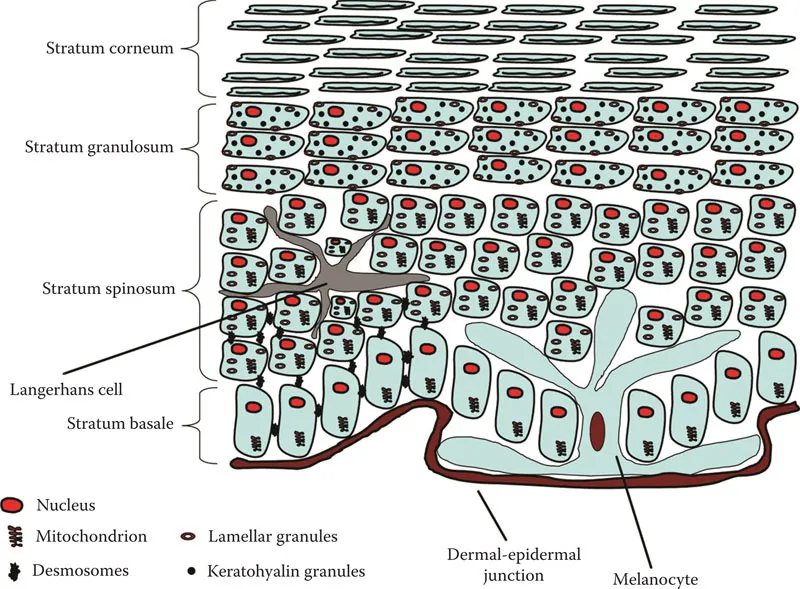
Figure 1.2 Diagram of the epidermal strata of skin.
Stratum basale
The stratum basale (or stratum germinativum) is the lowest layer in the epidermis and consists of a single layer of cells, which are predominantly keratinocytes. In this part of the epidermis, the keratinocytes are undifferentiated and contain all of the usual organelles that are present in viable cells, such as mitochondrion, Golgi apparatus, ribosome, endoplasmic reticulum, lysosome, and nucleus. The stratum basale is germinative and contains several populations of keratinocytes: stem cells, transient amplifying cells, and postmitotic cells. Stem cells divide rather infrequently and give rise to daughter cells known as transient amplifying cells. In turn, transient amplifying cells divide much more frequently than stem cells, producing postmitotic cells—the cells that actually undergo differentiation.
Keratinocyte cells are joined together by junctions known as desmosomes and are anchored to the underlying dermal-epidermal junction by hemidesmosomes. Desmosomes and hemidesmosomes are rather intricate structures composed of several macromolecules. The cytoskeleton of the keratinocytes consists of keratinocyte intermediate filaments, which provide structural support for the cell by traversing the cytosol. They are long, cylindrical proteins that are arranged concentrically around the nucleus and protrude to the limits of the cell—the plasma membrane—where they anchor to desmosomes and hemidesmosomes. Intermediate filaments anchored to hemidesmosomes are usually referred to as tonofilaments.
In the stratum basale, keratinocytes express K5 (58 kDa) and K14 (50 kDa), which form fine bundles and provide the cell with structural stability and flexibility. At the germinative layer, the cytoskeletal components must confer enough flexibility so that cell replication and division can take place.
Integrins are a family of receptor proteins that are found on keratinocytes in this layer. They participate in the attachment of cells to other cells and cells to extracellular matrix, that is, cell-to-cell and cell-to-extracellular matrix. They contain two subunits, alpha and beta. There are various forms of the alpha and beta subunits, which form many different types of integrins. Nevertheless, integrins play an important part in the stability/adhesion of the multicellular agglomerate of the epidermis.
In addition to keratinocytes, there are also melanocytes interspersed in this layer of the epidermis and to a lesser extent Merkel cells. Melanocytes produce the chromophore melanin, responsible for skin pigmentation, which form aggregates in cellular organelles called melanosomes. In turn, melanosomes are transferred from melanocytes to basal layer keratinocytes in order to protect its cellular components, specifically the chromosomes (DNA) in the nucleus. The Merkel cell, on the other hand, associates with dendritic endings of neurons forming a Merkel disc. The resulting complex (Merkel disc) is a mechanoreceptor; hence, it serves as a sensory receptor for light touch.
Stratum spinosum
In the spinous layer, there is a gradient of shapes adopted by the keratinocyte. At the lower level of the stratum spinosum, the cells have a structure more closely resembling that observed in the stratum basale; however, progressively closer to the stratum granulosum the cells become flatter. The stratum spinosum is extremely rich in desmosomes, providing multiple linkages between the keratinocytes. When the skin is prepared for histological studies, the cells shrink and the connections made by the desmosomes are readily visible, revealing the spinous appearance of this tissue, thus its name.
At this stage of differentiation, typically in cells located at higher levels of the stratum spinosum, we find the appearance of lamellar granules—secretory organelles that carry lipids and enzymes, which will be utilized for the formation of the stratum corneum. The organelles are about 0.2–0.3 μm in diameter, and contain phospholipids, sterols, glycoproteins, glycolipids, acid hydrolases (lipases, proteases, glycosidases, and acid phosphatase), and glucosylceramide.1
Residual K5 and K14 from the stratum basale still remain in the keratinocytes, although newly synthesized K1 and K10 appear in the stratum spinosum—forming larger bundles of intermediate filaments in this layer.
Langerhans cells, star-shaped in structure, are most abundant in this layer of the epidermis. These cells are essentially macrophages, derived from bone marrow, which migrate to the epidermis where they act as outposts of the immune system. They are antigen-presenting cells (APCs), which means they can patrol the epidermis to captu...
Table of contents
- Cover
- Half Title
- Title Page
- Copyright Page
- Dedication
- Table of Contents
- Preface
- Acknowledgments
- 1 Structure and function of skin
- 2 Free radicals in biology
- 3 The skin’s endogenous antioxidant network
- 4 Effects of solar radiation, air pollution, and artificial blue light on the skin
- 5 Lipid peroxidation and its measurement
- 6 Antioxidant assays
- 7 Electron spin resonance of skin
- 8 Treatment of skin with antioxidants
- 9 Antioxidant properties and application information
- Appendix 1: Glossary of terms
- Appendix 2: Biologically important molecules and mechanisms
- Appendix 3: Cellular signaling in skin
- Appendix 4: Thermodynamic and kinetic factors that contribute to antioxidant behavior
- Index
Frequently asked questions
Yes, you can cancel anytime from the Subscription tab in your account settings on the Perlego website. Your subscription will stay active until the end of your current billing period. Learn how to cancel your subscription
No, books cannot be downloaded as external files, such as PDFs, for use outside of Perlego. However, you can download books within the Perlego app for offline reading on mobile or tablet. Learn how to download books offline
Perlego offers two plans: Essential and Complete
- Essential is ideal for learners and professionals who enjoy exploring a wide range of subjects. Access the Essential Library with 800,000+ trusted titles and best-sellers across business, personal growth, and the humanities. Includes unlimited reading time and Standard Read Aloud voice.
- Complete: Perfect for advanced learners and researchers needing full, unrestricted access. Unlock 1.5M+ books across hundreds of subjects, including academic and specialized titles. The Complete Plan also includes advanced features like Premium Read Aloud and Research Assistant.
We are an online textbook subscription service, where you can get access to an entire online library for less than the price of a single book per month. With over 1.5 million books across 990+ topics, we’ve got you covered! Learn about our mission
Look out for the read-aloud symbol on your next book to see if you can listen to it. The read-aloud tool reads text aloud for you, highlighting the text as it is being read. You can pause it, speed it up and slow it down. Learn more about Read Aloud
Yes! You can use the Perlego app on both iOS and Android devices to read anytime, anywhere — even offline. Perfect for commutes or when you’re on the go.
Please note we cannot support devices running on iOS 13 and Android 7 or earlier. Learn more about using the app
Please note we cannot support devices running on iOS 13 and Android 7 or earlier. Learn more about using the app
Yes, you can access Antioxidants and the Skin by Roger L. McMullen in PDF and/or ePUB format, as well as other popular books in Medicine & Biochemistry in Medicine. We have over 1.5 million books available in our catalogue for you to explore.