Cardiovascular disease remains the chief cause of mortality and morbidity in adults in many parts of the world, and diagnosis and treatment is increasingly based on cellular, intracellular, and molecular parameters as well as systems analysis. Consequently, it is vital that medical students learn the fundamental physiology of the cardiovascular system. This book, along with its interactive electronic learning modules, breathes life into the subject, with animations, videos, and game-like decision-making.
- 152 pages
- English
- ePUB (mobile friendly)
- Available on iOS & Android
eBook - ePub
About this book
Trusted by 375,005 students
Access to over 1.5 million titles for a fair monthly price.
Study more efficiently using our study tools.
Information
Topic
MedicineSubtopic
CardiologySECTION II |
| CARDIOVASCULAR SYSTEM |
About 5 liters of blood per minute circulate around your body even when you are resting. In other words, 5 one-liter soft drink bottles full of blood moved every 60 seconds! During exercise that amount can increase to 20 liters per minute or more! Moved from where to where? At any given moment, most of the blood in your body, 60% to 70%, resides in the veins. A major function of the heart is to receive blood at low pressure from the venous compartment, transport it through the lungs, and increase the pressure as the blood is ejected into the arterial conduits. The blood then flows from the high pressure arterial side of the circulation back to the low pressure venous side, and so on, continuously in a circle, just as demonstrated first by William Harvey in the seventeenth century.
What does the above have to do with the pathophysiology of heart disease? Just this—the body’s demand for nutrition supplied by blood flow continues unabated when ventricular function becomes abnormal. Overall body metabolism persists, the demand of the body for oxygen continues, and the body’s feedback mechanisms strive to maintain blood flow into the microcirculation for gas and other exchange. The feedback mechanisms act mostly via the autonomic nerves to the heart, blood vessels, and adrenal gland. Quite simply, the body has no mechanisms to rest a weakening heart. There is a continuing drive to produce cardiac output adequate for body metabolism. Moreover, this drive continues in the presence of abnormal heart valves or heart muscle pathology. These continuing demands on the abnormal heart lead to ventricular dysfunction, adverse myocardial myocyte remodeling, and the symptoms we diagnose as heart disease.
As you can understand from the above, one cannot diagnose and treat heart disease without understanding how the normal circulation works, including some of the simple physics of circulating fluid, how the heart pumps blood, and the characteristics of the peripheral circulation, controls in the circulation, individual organ circulation, and the microcirculation.
8 |
| How the circulation works |
Blood pressure
A little physics is useful here. Pressure can be measured as the height of a column of fluid connected to a pressurized “container,” such as a blood vessel (Figure 8.1). Use of the height of a fluid column is based on pressure = ρgh; ρ is fluid density, g is acceleration due to gravity, and h is the height of the fluid column.
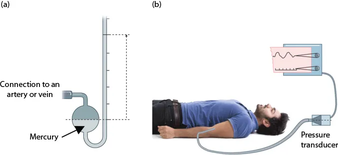
Figure 8.1Blood pressure measuring devices. (a) Mercury-filled U-tube; (b) electronic pressure transducer. Pressure is measured in a hospital catheterization laboratory or intensive care unit with electronic pressure transducers as in (b) The tubing from an artery or vein (b) is filled with sterile saline. Blood pressure within, for instance, an artery pushes against the saline column in the tubing and the pressure is transmitted to the pressure transducer (b). A pressure transducer (b) transforms pressure into voltage that is easily displayed and measured (b). Direct connection to a mercury-filled system (a) has been used in the past, but is avoided because of concerns about mercury toxicity. Most clinical pressure measurement devices are spring-loaded or electronic sphygmomanometers. They are smaller and more convenient to use than a fluid column. Any pressure-measuring device can be called a manometer and “sphygmo-” refers to pulse. Blood pressure measuring devices are calibrated with a mercury column (a).
The density, ρ, of a fluid such as mercury or water and the acceleration due to gravity, g, are known and constant, so pressure can be expressed simply as the height (h) of a column of liquid. Two basic types of pressure measuring devices are illustrated (Figure 8.1a and b).
Energy
Most of the energy in the circulation ultimately derives from the hydrolysis of ATP in heart muscle and conversion of high energy phosphate bonds by sarcomeres into heart muscle work. Work is done by heart muscle on blood contained within the heart and that work is transformed into two types of energy: pressure potential energy and energy related to movement, kinetic energy. A third type of energy is not derived from heart muscle function. It is derived from the effects of gravity on blood in the circulation. Energy in the circulation will be defined first and then flow will be discussed, including a discussion of gravity’s effects on the circulation.
Total fluid energy is defined as follows:

P, pressure potential energy, develops due to the work done on the blood in the ventricle by the contracting heart muscle myocytes during systole. Contracting ventricular muscle squeezes blood within the ventricular chambers, which cannot decrease in volume because the valves are all closed. Blood, like water or saline, is incompressible at in vivo pressures. Pressure builds up in the blood in the ventricular chambers and when the pressure reaches the pressure level in the pulmonary artery and aorta, the respective valves are pushed open. The ventricles then push the blood into the great vessels, which already contain pressurized blood. This forces the vessels to expand and aortic and pulmonary artery pressure to increase further.
Kinetic energy, (ρv2)/2, appears because some pressure potential energy is transformed into movement of the blood through the great vessels; v, velocity, is distance moved per unit time. Density, ρ, is used to refer all calculations to a unit volume and mass of blood. In a person at rest, kinetic energy in the circulation relative to the other terms is small, approximately 3% to 5% of total energy, b...
Table of contents
- Cover
- Half Title
- Title Page
- Copyright Page
- Contents
- Preface
- Goals
- SECTION I: CARDIAC ELECTROPHYSIOLOGY AND THE ELECTROCARDIOGRAM (ECG)
- SECTION II: CARDIOVASCULAR SYSTEM
- References for additional reading
- Index
Frequently asked questions
Yes, you can cancel anytime from the Subscription tab in your account settings on the Perlego website. Your subscription will stay active until the end of your current billing period. Learn how to cancel your subscription
No, books cannot be downloaded as external files, such as PDFs, for use outside of Perlego. However, you can download books within the Perlego app for offline reading on mobile or tablet. Learn how to download books offline
Perlego offers two plans: Essential and Complete
- Essential is ideal for learners and professionals who enjoy exploring a wide range of subjects. Access the Essential Library with 800,000+ trusted titles and best-sellers across business, personal growth, and the humanities. Includes unlimited reading time and Standard Read Aloud voice.
- Complete: Perfect for advanced learners and researchers needing full, unrestricted access. Unlock 1.5M+ books across hundreds of subjects, including academic and specialized titles. The Complete Plan also includes advanced features like Premium Read Aloud and Research Assistant.
We are an online textbook subscription service, where you can get access to an entire online library for less than the price of a single book per month. With over 1.5 million books across 990+ topics, we’ve got you covered! Learn about our mission
Look out for the read-aloud symbol on your next book to see if you can listen to it. The read-aloud tool reads text aloud for you, highlighting the text as it is being read. You can pause it, speed it up and slow it down. Learn more about Read Aloud
Yes! You can use the Perlego app on both iOS and Android devices to read anytime, anywhere — even offline. Perfect for commutes or when you’re on the go.
Please note we cannot support devices running on iOS 13 and Android 7 or earlier. Learn more about using the app
Please note we cannot support devices running on iOS 13 and Android 7 or earlier. Learn more about using the app
Yes, you can access Cardiovascular Physiology by Burt B. Hamrell in PDF and/or ePUB format, as well as other popular books in Medicine & Cardiology. We have over 1.5 million books available in our catalogue for you to explore.