New neuroimaging techniques are developing at a break neck pace-every academic journal contains glossy pictures of brain activity corresponding to a particular task emblazoned in glorious technicolor. Discoveries about brain function in psychiatric disorders have been made at an equally rapid rate. However, most books on the subject have been writt
Trusted by 375,005 students
Access to over 1.5 million titles for a fair monthly price.
1. FUNCTIONAL NEUROIMAGING: AN INTRODUCTION TO THE TECHNOLOGY, METHODOLOGY, INTERPRETATION, AND APPLICATIONS
Tamara A Russell, Fernando Zelaya, Rodrigo A Bressan, Peter A Bandettini
Introduction
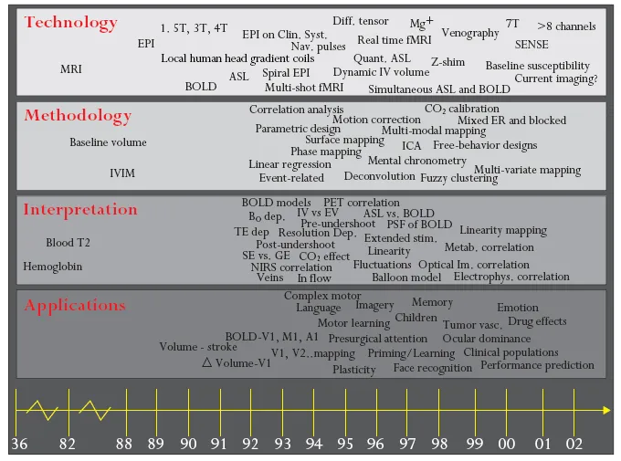
Many technological advances have been seen in the neuroimaging field. These advances are allowing the investigation of new and exciting neurophysiological and pathophysiological questions. This chapter intends to provide the reader with a brief overview of the principles of MRI, fMRI, PET and SPET. Basic information on the hardware, specific applications and major limitations of the different neuroimaging techniques is provided to give the reader a perspective of what can and cannot be done with these techniques. Functional magnetic resonance imaging (fMRI) has been in existence for about 11 years.1–3 During this time, the technique has experienced explosive growth. The reasons for this growth include the following: minimal invasiveness; the growing availability of the necessary hardware; the unique functional spatial and temporal resolution niche that it fills; its ability to map a network of substrates that intervenes in cognitive and sensory processes; and, importantly, the potential that it promises for the investigation of abnormal brain function. Figure 1.1 is an attempt to graphically illustrate a timeline of the advancement of many, but certainly not all, significant aspects of functional imaging technologies.
Although the use of this tool has increased exponentially, it is still quite difficult for the average end-user, perhaps a psychologist or psychiatrist, to come to grips with the basic principles and physics of MR. The aim of the first section of this chapter is to provide an overview of these basic principles. Subsequent sections will describe aspects of the hardware used in the MRI environment, and advances and developments that are being made in this area. Additionally, some discussion of the neuronal information extracted from the blood oxygenation-level-dependent (BOLD) signal will be discussed. Lastly, the applications of this tool are briefly outlined.
Figure 1.1 Growth and progression of neuroimaging techniques. A1, auditory cortex; ASL, arterial spin labeling; B0, external field; BOLD, blood oxygenation-level dependent; EPI, echo planar imaging; ER, event related; EV, extravascular; fMRI, functional magnetic resonance imaging; GE, gradient echo; ICA, independent component analysis; IV, intravenous; IVIM, intravoxel incoherent motion; IV v EV, intravascular versus extravascular; M1, motor cortex; MRI, magnetic resonance imaging; NIRS, near infrared spectroscopy; PSF, point spread function; SE, spin echo; SENSE, sensitivity encoding for fast MRI; T, tesla; V1, visual area 1; V2, visual area 2.
Basic principles of functional magnetic resonance imaging (fMRI)
fMRI is a new application of an existing technology (i.e. MRI). This section will attempt to introduce, in a basic way accessible to the novice, the elements involved in the production of the MRI signal. It will cover basic principles relating to the nuclei of hydrogen atoms (which is where most of the MR signal comes from) and their properties when an external magnetic field is applied. Precession and resonance will be described, as will the mechanisms by which protons can be excited and relaxed. By necessity, in order to provide a brief introduction to this area, some aspects of this explanation may be deemed by the more experienced reader as oversimplified. It is stressed at this point that the purpose of this chapter is to provide an introductory overview to the novice neuroimager, providing a level of understanding that will demystify some of the principles and terminology used, not to provide an in-depth understanding of all the physics involved in MRI.
Nucleus of the hydrogen atom
Hydrogen atoms give rise to most of the signal in an MR image. The nucleus of the hydrogen atom consists of one proton and no neutrons. This gives the hydrogen atom a positive charge and an atomic number of 1, as there is only one proton in its nucleus. Hydrogen atoms are abundant in the human body: approximately 70% of the body is made up of water (containing two hydrogen atoms and one oxygen atom). In the brain, gray matter has approximately a 70% water content, blood approximately a 93% water content and white matter (glial cells) approximately an 85% water content. The large quantity of hydrogen atoms in the human body and the large magnetic moment (see below) of the single proton in the nucleus of the atom are responsible for the large MR signal they produce when compared with those of other nuclei.
Magnetic moment
Some atomic nuclei have the property of ‘spin’. This means that they have a net angular momentum, i.e. they can be seen as ‘rotating’ around their main axis. The spinning protons combined with their net electric charge create what is called a net magnetic moment, as if they were a small magnet with a north and a south pole.
External magnetic fields
In the absence of any external magnetic field, proton magnetic moments are randomly orientated and (as a group) are considered to have a net magnetization of zero. When an external magnetic field is applied to an object, the spin axes of all the nuclei in the object line up with the magnetic field. The nuclei can either align parallel to the magnetic field or anti-parallel to it (i.e. in the opposite direction). Factors which influence the direction of orientation include the thermal energy of the atoms and the strength of the external magnetic field. High-energy protons are strong enough to be able to align themselves anti-parallel to the external magnetic field while those with low energy will align in a direction parallel to the magnetic field. In reality, the number of protons that align parallel and anti-parallel with the field is not the same. This difference produces a net magnetization of the whole sample (in our case, the human brain). With increases in magnetic field strength, this difference increases, therefore enhancing the net magnetization of the sample.
For simplicity, it is easier to consider from here onwards only the net magnetization of the sample. As you may have realized, this net magnetization has a definite orientation, i.e. in the direction of the excess of protons that align parallel with the field. Therefore, it is represented as a vector quantity, which is termed the net magnetization vector (NMV) or M.
Precession
Because of their intrinsic angular momentum (described above), protons do not align with the external field (parallel or anti-parallel) in a ‘straight’ line. Instead, they behave just like a spinning top does as it starts to fall. Precession is the term used to describe the way in which protons ‘wobble’ around their axes. When an external magnetic field is applied, the protons precess in line with it, but wobble in a conical manner that is similar to the spinning top rotating around a vertical axis. Precessional frequency is a term used to describe the rate (or speed) at which the protons or the NMV precesses.
Resonance and the Larmor frequency
When exposed to an external magnetic field, all hydrogen protons will precess at the same frequency. This frequency is determined by the gyromagnetic ratio of the particular protons and the strength of the magnetic field. It is described by a very simple equation called the Larmor equation, which states that the frequency of precession (
) is given by the product of the gyromagnetic ratio of the protons (
) times the strength of the external field (B0):
The gyromagnetic ratio of hydrogen is 42.57 MHz/tesla (T) and all hydrogen protons will precess at this same frequency when exposed to an external magnetic field of 1 T. The strength of the magnetic field (from the scanner) is measured in tesla (T) or gauss (G): 1 T is equivalent to 1 × 10⁴ G (about 20,000 times stronger than the earth’s magnetic field).
As can be seen, this equation determines the frequency at which the protons will resonate. The resonant frequency is also referred to as the precessional frequency or the Larmor frequency. The equation also demonstrates that the higher the magnetic field, the greater the precessional frequency.
The strength of the magnetic field will usually increase the intensity of the signal because the net magnetization will be larger. It is also worth noting that fMRI contrast also increases approximately linearly with field strength.4–6 Field strength drives both the frequency of precession of protons and the fMRI contrast in a linear fashion. The drive for an increased signal:noise ratio (S:N; i.e. the height of the signal of interest compared to the background noise) and for increased functional contrast has been respons ible for the proliferation of higher field strength magnets for human fMRI, from typically 1.0 T in 1984 to the present day where scanners in the range of 4–7 T are being developed for human use. Field strengths as high as 12 T are being developed for animal use. The majority of scanners in use in academic centers are in the range of 1.5–4 T. Higher field magnets cost more to build and maintain, and create their own technical hurdles. Some of the advantages and disadvantages of high field strength are outlined in Table 1.1.
But what actually is resonance? Resonance refers to the property of a body (in this case the nucleus of an atom) to absorb energy at a characteristic, natural frequency. In MR, the natural frequency for resonance absorption is given by the Larmor equation. A condition of resonance can be achieved by placing the protons within a strong external field and exciting them with a second, alternating magnetic field [in the form of radiofrequency (RF) waves, see below]. When resonance occurs, not only do the magnetic moments of the protons change their angle of rotation, but all the protons begin to precess in phase with each other (referred to as phase coherence).
Protons can absorb energy from external sources but also give off energy when they try to change their alignment back to their initial configuration in the magnetic field. These two types of processes form the basis of how the MR signal is created and how it disappears.
Noll7 has described the resonance phenomena using the analogy of a guitar string. With a guitar, the frequency varies according to how much tension is applied to the string by the fingers [similar to the strength of the magnetic field (B0)]. The string is most often plucked in order to excite it and create resonance (in the form of acoustic waves), however, it can also be excited by holding up a loudspeaker near to the string and playing a note at the same frequency as the string. This is similar in manner to the way in which nuclear spins are excited by the application of RF waves (a second, alternating magnetic field of the correct frequency).
Table 1.1 Advantages and disadvantages of high field strength functional magnetic resonance imaging (fMRI)
The term excitation is used to describe the delivery of energy to the protons at their characteristic frequency. As the proton resonates it moves out of alignment with the external magnetic field (B0), to attempt to align with the second, alternating magnetic field. The NMV is now also changing out of alignment with B0. This alternating ...
Table of contents
COVER PAGE
TITLE PAGE
COPYRIGHT PAGE
CONTRIBUTORS
PREFACE
1. FUNCTIONAL NEUROIMAGING: AN INTRODUCTION TO THE TECHNOLOGY, METHODOLOGY, INTERPRETATION, AND APPLICATIONS
2. UPDATE ON FUNCTIONAL NEUROIMAGING IN CHILD PSYCHIATRY
3. NEUROIMAGING AND AGING
4. NEUROIMAGING STUDIES OF HUMAN DRUG ADDICTION
5. NEUROIMAGING STUDIES OF MOOD DISORDERS
6. NEUROIMAGING AND EATING DISORDERS
7. NEUROIMAGING AND THE PATHOPHYSIOLOGY OF OBSESSIVE-COMPULSIVE DISORDER (OCD)
8. IMAGING BRAIN STRUCTURE AND FUNCTION IN SCHIZOPHRENIA: NEW TECHNIQUES ENCOUNTER OLD PROBLEMS
9. FUNCTIONAL IMAGING STUDIES OF PSYCHOPATHY, ANTISOCIAL PERSONALITY DISORDER (APD) AND RELATED PSYCHOLOGICAL PROCESSES
Frequently asked questions
Yes, you can cancel anytime from the Subscription tab in your account settings on the Perlego website. Your subscription will stay active until the end of your current billing period. Learn how to cancel your subscription
No, books cannot be downloaded as external files, such as PDFs, for use outside of Perlego. However, you can download books within the Perlego app for offline reading on mobile or tablet. Learn how to download books offline
Perlego offers two plans: Essential and Complete
Essential is ideal for learners and professionals who enjoy exploring a wide range of subjects. Access the Essential Library with 800,000+ trusted titles and best-sellers across business, personal growth, and the humanities. Includes unlimited reading time and Standard Read Aloud voice.
Complete: Perfect for advanced learners and researchers needing full, unrestricted access. Unlock 1.5M+ books across hundreds of subjects, including academic and specialized titles. The Complete Plan also includes advanced features like Premium Read Aloud and Research Assistant.
Both plans are available with monthly, semester, or annual billing cycles.
We are an online textbook subscription service, where you can get access to an entire online library for less than the price of a single book per month. With over 1.5 million books across 990+ topics, we’ve got you covered! Learn about our mission
Look out for the read-aloud symbol on your next book to see if you can listen to it. The read-aloud tool reads text aloud for you, highlighting the text as it is being read. You can pause it, speed it up and slow it down. Learn more about Read Aloud
Yes! You can use the Perlego app on both iOS and Android devices to read anytime, anywhere — even offline. Perfect for commutes or when you’re on the go. Please note we cannot support devices running on iOS 13 and Android 7 or earlier. Learn more about using the app
Yes, you can access Neuroimaging in Psychiatry by Cynthia H. Y. Fu, Carl Senior, Tamara Russell, Daniel R. Weinberger, Robin Murray, Cynthia H. Y. Fu,Carl Senior,Tamara Russell,Daniel R. Weinberger,Robin Murray in PDF and/or ePUB format, as well as other popular books in Psychology & Neurology. We have over 1.5 million books available in our catalogue for you to explore.