19.1.1 Our approach (USA)
Andrew Salzberg and Jordan Jacobs
The evolution in surgical treatment of breast cancer from Halsted’s radical mastectomy to the present day skin-sparing and nipple-sparing techniques has provided the reconstructive surgeon with more options.1,2,3 The resultant larger skin envelope is able to support immediate breast reconstruction, provided skin flaps are healthy.4,5 The single-stage, direct-to-implant approach overcomes the limitations of a two-stage, expander approach and represents the ultimate simplicity in breast reconstruction.
The feasibility of a single-stage implant approach has been greatly facilitated by the introduction of acellular dermal matrices (ADMs) in reconstructive breast surgery.6 These structurally intact tissue matrices provide the biologic scaffold necessary for tissue in-growth and cellular repopulation. They have been utilized to specifically provide coverage of the implant or expander at the inferolateral pole,7,8,9,10,11,12,13,14 thus eliminating the need for recruitment of neighboring muscle and/or fascia for this purpose.10 ADM use at the inferolateral pole also allows better control of the inframammary fold, improves lower pole projection, and reduces expander or implant migration.8,10 Collectively, these benefits of ADM can lead to improved aesthetic results.10,15
Preoperative assessment to determine whether the patient is a candidate for direct-to-implant reconstruction focuses on the patient’s body habitus, the ablative component of the surgery, and the specific desires of the patient for contralateral breast symmetry. Coordination between plastic surgeon and surgical oncologist will help ensure a clear understanding of the planned surgical strategy. The ideal candidate for a direct-to-implant technique is a patient with a medium or small breast size, grade 1–2 ptosis, and good skin quality. Patients with a history of smoking must abstain for at least 4 weeks prior to and after surgery. Morbidly obese patients are generally poor candidates for implant reconstruction because of an increased risk of postoperative complications.16
Although the quality of the skin flaps after mastectomy is a critical component for a successful outcome, choosing an appropriately sized implant to fill the space beneath these flaps is also an important consideration. The patient’s chest wall dimensions are carefully measured and an array of sizers is used preoperatively to gauge the anticipated volume that will be required. In addition, a three-dimensional volumetric computer program is commonly used to assist with implant selection. A notable point is to have an implant base width that complements the chest diameter, as a concave lateral chest contour will result if the implant is of insufficient width. Saline implants are not routinely recommended because they often produce a suboptimal aesthetic shape in a reconstructed breast and are prone to greater visibility and palpability as well.
MASTECTOMY AND RECONSTRUCTION
The reconstructive surgeon needs to be an active participant during the mastectomy, thereby assuring careful handling of the skin and the avoidance of traction or thermal injury to the flaps. If electrocautery is used for flap elevation, low settings are monitored and used on low cutting to reduce the risk of tissue damage. Scalpel or more recent radiofrequency devices (Peak™ Surgical, Palo Alto, CA) may also be used for surgical development of skin flaps.
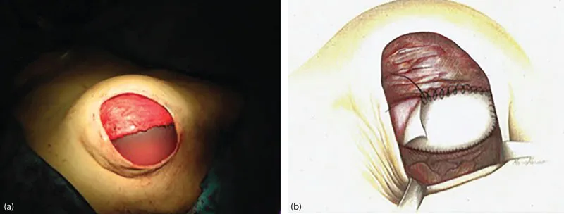
Critical for immediate reconstruction with a direct-to-implant technique is utilizing ADM to extend the submuscular plane, support the implant in its anatomic position, and define the inferior and lateral folds of the breast. The technique begins by creating a subpectoral pocket extending from the second rib superiorly, medially to the origin of the pectoralis major muscle at the sternum, and laterally to the lateral mammary fold. Elevation of a portion of the infero-medial muscle to allow anatomic placement of the implant is also performed. The ADM is introduced at the inferior pole (Figure 19.1.1.1) and is sutured in a running fashion to the pectoralis major muscle along its entire lower course from medial to lateral and then around and down to the lateral mammary fold.
No elevation of muscle is necessary at the serratus margin and direct suturing of the ADM to the chest wall with absorbable suture material defines and sets the limit of the lateral fold. The appropriately sized implant is confirmed by surgical judgment as well as by the weight of the mastectomy specimen. A modest over correction is added to the final implant volume to accommodate the anticipated release of the skin elasticity following mastectomy. After placing the implant beneath the muscle-ADM layer, continued suturing of the ADM directly to the inframammary fold at its desired position—without expectation of migration—is performed, completely covering the implant. Direct conformation of the skin envelope to the lower pole will decrease the chance of seroma formation. A “perfect” shape should be achieved at this point and postoperative settling should not be planned. Two suction drains are placed in the subcutaneous plane through separate stab incisions followed by skin closure.
Typically, patients remain in hospital for approximately 48 hours. Upon discharge, drains covered with occlusive dressings are maintained for 7–14 days, with removal determined by the amount and quality of their output. A supportive surgical bra with an additional superior pole pectoralis strap is worn for a minimum of 3 weeks to assist in the optimal positioning of the implants in the pocket. The patients are encouraged to do gentle exercise to improve range-of-motion of the arm and to perform gentle massaging of their breasts to prevent development of axillary contracture.
This technique can be successfully performed in patients with a variety of breast sizes and varying degrees of ptosis. Moreover, these techniques are suitable for patients undergoing nipple-sparing or skin-sparing mastectomy for either oncologic or prophylactic reasons. The principles for clinical decision making in the context of single-stage implant reconstruction with ADM are summarized in Figure 19.1.1.2.
Over a 14-year period, low rates of complications and good aesthetic outcomes (Figures 19.1.1.3 and 19.1.1.4) have been consistently obtained. Major complications include implant loss (1.4%), skin necrosis requiring re-operation (1.1%), infection (0.9%), hematoma (0.6%), seroma (0.5%), and capsular contracture (0.5%). Other minor issues related to suture exposure, wound healing delay, superficial epidermolysis, and non-infectious redness of the skin have been noted in up to 1% of patients.

