Ophthalmic Imaging serves as a reference for the practicing ophthalmic imager. Ophthalmic imaging combines photography and diagnostic imaging to provide insight into not only the health of the eye, but also the health of the human body as a whole. Ophthalmic photographers are specialists in imaging through and in the human eye, one of the only parts of the body where the circulation and nervous system is visible non-invasively. With technical perspective as context, this book will provide instructional techniques as well as the background needed for problem solving in this exciting field. The book covers all aspects of contemporary ophthalmic imaging and provides image support to ophthalmologists and sub-specialties including retinal specialists, corneal specialists, neuro-ophthalmologists, and ocular oncologists. This text serves as a reference for the practicing ophthalmic imager, or to imagers just getting started in the field.
eBook - ePub
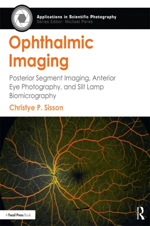
Ophthalmic Imaging
Posterior Segment Imaging, Anterior Eye Photography, and Slit Lamp Biomicrography
- 374 pages
- English
- ePUB (mobile friendly)
- Available on iOS & Android
eBook - ePub
Ophthalmic Imaging
Posterior Segment Imaging, Anterior Eye Photography, and Slit Lamp Biomicrography
About this book
Trusted by 375,005 students
Access to over 1.5 million titles for a fair monthly price.
Study more efficiently using our study tools.
Information
Topic
MedicineSubtopic
Digital MediaPart 1
Introduction
What Is Ophthalmic Photography and Imaging?
Technology allows remarkable access to the inner world of the human body. Through medical imaging, we can see bones and tissues inside the body, watch the brain “light up” as it performs in a variety of ways, or visualize the blood flow as it makes its way through an organ. The primary goal of many of these images is showing us things we cannot otherwise see. All these methods act as a window into the structure and function of the body and give us insight into what may be going wrong.
However, the term “photography” rarely comes up in this context. There’s a good reason for that; photographs capture a moment in time, but they do not often show us what we don’t already know is there. Photography is a great tool for documenting what the patient looks like on this particular day and time. But in order for information to be diagnostic, it needs to provide insight above and beyond observational information. Other forms of medical imaging, such as magnetic resonance imaging (MRI) or x-rays, provide a view of the body not otherwise seen, allowing a physician to gain additional information that could point to a specific diagnosis. In other words, photography is generally considered to be documentary, and medical imaging diagnostic.
Ophthalmic photography and imaging is the practice of capturing photographs or other images of the eye. The more important part of that definition is why these images are made; they are captured in direct support of a patient’s medical care. We image eyes for a number of reasons in ophthalmology. These include documenting what someone’s eyes look like at a particular moment in time to compare over subsequent visits, using photographs to illustrate a particular disease process for both documentation and education, and even to provide diagnostic information to the physician. Ophthalmic photography holds a unique distinction of being the only type of photography that has diagnostic capability.
You may want to stop me here, and say, “What about x-rays, CT, MRI?” Again, the distinction lies in the term “photography.” As mentioned, photography with an ordinary camera is often considered to be a documentary device—what you see is what you get (mostly). In other words, photography as a tool is not going to radically change what you are seeing with your own eyes. (For the purposes of this technically focused discussion, we are setting aside for now the use of photography as a conceptual art medium.)
In its most common applications, photography uses visible spectrum energy to expose a light sensing material; film, or more commonly these days, digital sensor technology. This means that photography uses the same light energy and color distinctions that we see with our own eyes; again, this reinforces the idea of what you see is what you get. In fact, photography is designed specifically to mimic the human visual response so that photographs look as we expect them to. All other medical imaging modalities, including x-rays, MRI, CT scans and even ultrasounds are not photography. These imaging techniques use varying parts of the electromagnetic spectrum to penetrate into the body, which in turn are translated into images. So while photography and imaging are cousins, as they both use portions of the electromagnetic spectrum, the term “photography” connotes something very different that is separate from medical imaging.
In fact, the thing that allows ophthalmic photography to be diagnostic has very little to do with photographic capability. The key to what makes ophthalmic photography so special lies not in the cameras it is captured with (they are just cameras) but how we generate the images of the eye in the first place.
The eye is a pretty special organ for many reasons. It is an incredibly complex structure with many of the key imaging components visible simply by looking in the mirror. That means we can learn a lot by looking carefully at the parts of the eye we can see, as long as we know what we are looking at or for. This makes the eye a great subject for photography to document anything that might be out of the ordinary; the only special equipment we need to make photographs of those structures is a close-up (macro) lens and a bright enough light source.
For the parts of the eye we cannot see easily, such as the retina, we just need to pharmaceutically dilate the pupil and shine a light inside. (We get a hint of how easy this is to do from red-eye photos; the red-eye phenomenon is caused by the reflection of the light source in the retina.) Once dilated, we can see the retina (inner lining of the eye), which provides a window into not only a patient’s ocular health, but also the health of the nervous system and cardiovascular system. Since these structures are visible with dilation (with some help from specialized optics and illumination), ophthalmic photography takes this access a step further with angiography, or dye testing. Angiography takes ophthalmic photography from documentation to diagnosis by providing the ophthalmologist with an illuminated view of the retinal vessels as the dye travels through them.
The saying about the eyes as a window to the soul is pretty spot on, at least from a medical point of view. The eye is the only part of the body in which we can visualize so many systems without cutting anything open. The non-invasive, diagnostic potential of this type of imaging is only just being realized for systemic disease screening and rural patient out-reach. (We will touch on the applications of telemedicine in this book too.) Diabetes, high blood pressure, some cancers and other destructive systemic diseases are all visible in the retina without angiography or dye injection; there is even research emerging on the early detection of Alzheimer’s and other brain diseases via retinal imaging.
The founding technologies of ophthalmic photography, fundus photography and fluorescein angiography are photographic techniques. They were devised and practiced for many years using film, which required exposing, processing and printing the images in a darkroom. Visible light was and is still used to make exposures of the eye, though some modern techniques employ energies just outside the visible spectrum as well.
In the early years of the field, photographers from other disciplines were hired into ophthalmic photography positions, working for doctors and hospitals. These photographers processed retinal images captured on film in the darkroom and printed them into positive images from the negatives. They were trained to perform ophthalmic photographs on the job, in addition to learning about the anatomy of the eye and ophthalmic disease. Even more important than technical skills in the darkroom, photographers’ innate understanding of light and its behaviors, as well as optics and image formation knowledge, proved extremely useful and complementary to the ophthalmic field.
Fast forward 40 years or so and fundus photos and fluorescein angiograms are still done, but they are now captured using digital cameras. Digital imaging expanded the capability of the ophthalmic imager to include other techniques, such as indocyanine green angiography (ICG) and fundus autofluorescence imaging. In fact, the very nature of digital imaging has transformed imaging into measurable, quantified data in ways that were never possible with film. We have added other imaging types to our repertoire, such as optical coherence tomography, that have little to do with photographic principles. Technologies that incorporate light and optical principles are a natural fit with the skills and background of an ophthalmic imager.
Having said all of that, many folks in the field still call themselves “ophthalmic photographers” as a nod to those early days. Regardless, these individuals have a unique combination of skills: knowledge of light and its behaviors in imaging, combined with a working knowledge of eye diseases and how to illustrate them through images. These images can be technical in nature, but they work to establish a complete visual narrative that is descriptive of the patient’s condition. “Photographer” in this case is less about how the image is created, as it is about what that person’s expertise can include. However, clinical environments and subsequent posted positions may use the terms “photographer” and “imager” interchangeably.
Getting back to the original premise of this discussion, you might notice the name of this book is not “Ophthalmic Photography,” but “Ophthalmic Imaging.” This is precisely because the field has expanded to include not only photographic techniques, but imaging modalities such as optical coherence tomography that have more in common with ultra-sounds, CT scans or confocal imaging than they do with photography. The term “imager” is more descriptive of the comprehensive techniques and technologies utilized in modern ophthalmology.
This is an important distinction; for our purposes and throughout the book, the term “imaging” includes any methodology used to create an image of the eye.
To be a successful ophthalmic imager, there is a need to learn the techniques and technology of ophthalmic imaging and also the desire to help patients in the most compassionate manner possible. Most of us take our good vision for granted; the very thought of losing vision is visceral and terrifying. Above all, patients are human beings facing what could be the biggest medical and psychological challenge they have ever encountered. As a part of the health care team, the ophthalmic imager must perform at the highest possible level, and should do so with empathy and compassion.
After all, after learning (and perfecting!) the techniques and technologies outlined in this book, the biggest challenge will always be your patients. Patients will present with physical challenges including various mobility impairments, optical challenges like cloudy media, or emotionally challenges as you struggle to figure out how to provide care despite how they (or you) feel. Patient care is not for everyone, but knowing you are putting forth your best effort to give that patient the optimum chance of saving their vision can be extremely gratifying.
It is my hope that this book will help you on your journey to learn more about ophthalmic imaging, both the field and the practice. This is an exciting, growing field, with many emotional rewards! I strongly suggest looking through the section “How to Use This Book” next to tailor the book to your specific needs.
How to Use This Book
There is no single or “right” path to becoming an ophthalmic imager. The most common denominators among practicing ophthalmic imagers are a passion for imaging and photography and a curiosity for all things related to the human eye. Most practicing ophthalmic imagers discover the field by chance, and many learn on the job. Unlike x-ray, ultrasound or MRI technicians, there is no degree in ophthalmic photography and imaging.
There are few places in the United States to gain formal training in this field. I am lucky enough to teach at one of them in my role here at the Rochester Institute of Technology, in the only course sequence in ophthalmic imaging offered as part of a bachelor’s degree in photographic sciences. However, the demand for trained individuals far outweighs the supply, and I am constantly hearing from people wanting to learn more about ophthalmic imaging, but for whom it does not make sense to earn a degree (or another degree) just to gain that knowledge.
This book is an effort to capture that information in an accessible way. While there is no substitute for hands-on learning, my hope is that this book will provide the background and information to complement clinical experience and on-the-job learning.
If you are just starting out with no imaging experience and no ophthalmology experience, read this book from cover to cover. Then, find out when and where the next meeting of the Ophthalmic Photographers’ Society (OPS) is (opsweb.org) is, because that is where you can take workshops and courses to apply these principles hands-on. You will also meet with people just like you, either experienced or just starting out in the field. The OPS is a fantastic resource for networking and career placement.
If you are already in the ophthalmology field and familiar with ocular anatomy, but are just beginning to learn the imaging side, focus more on the photography, photographic technology (“Foundations,” Part 2) and imaging chapters of the book. These are written to provide the background about how these devices work, which will allow you to troubleshoot your own images much more effectively. Manufacturers love to say that some of these devices are becoming easier and easier to use, which in some cases is true. But being a button pusher is not very fulfilling, and when something goes wrong, that person has no recourse other than to throw up their hands. Having insight into how these imaging modalities work provides you with the ability to troubleshoot and correct problems, which makes you a much more valued employee than the average button pusher. It also ensures the best possible patient care, because that expertise is backed up with knowledge about why those images are being done. Even if it is technically “easy” to take an image, you need to understand what you are taking an image of and why you are doing it in the first place. (More on that in Part 5 on Imaging Diseases)
Moreover, new technologies on the horizon are often anything but user-friendly, and they will fall squarely in the “imaging expert’s” wheelhouse. As an essential part of the ophthalmology healthcare team, the expert ophthalmic photographer stands shoulder to shoulder with ophthalmologists as they help decide whether the latest imaging modality is truly showing new information or if old information is just being presented in a new shiny package. Ophthalmic imagers also provide insight into patient workflow and are responsible for generating revenue; ophthalmic imaging techniques are among the most highly reimbursed activities in any ophthalmologist’s office. In other words, the more skilled and knowledgeable the imager is, the better off the practice is on any number of levels.
Finally, if you are someone with an interest or background in photography but are looking to learn everything you can about ophthalmic imaging, start with the anatomy chapter (“Anatomy of the Eye,” Chapter 1). This will provide you with the background information on the human eye as a subject. Then, make your way through the sections on imaging, especially the sections on imaging disease, as this will help provide context about why we take these images in the first place. It will also tell you about how different diseases “behave” under different imaging modalities.
If you are currently working in ophthalmology, you may not have a traditional fundus camera in your practice. You may have an optical coherence tomographer (OCT), a scanning laser ophthalmoscope (SLO), and/or a ultra-wide-field camera (UWF). It may be tempting to skip through the sections on the devices you do not have. However, I would strongly encourage you look at fundus photography and angiography in particular, since those sections provide an excellent starting point to learning about how all these images are taken and even provide the basis for how the rest of these devices work.
The last piece of the puzzle in becoming a skilled ophthalmic imager is practice. There is no greater learning experience than dealing with patients every day, each with different eyes, different diseases and different challenges. Immersion in that environment leaves you no choice but to become better at capturing images. Be proactive! Ask questions of your doctors, such as: why did you order this image series/type for this disease process? What do the images tell you about this disease? How could the images be made better? Gain feedback and (constructive) criticism; this is by far the best way to improve and ensure your images are meeting their needs.
In a busy clinical environment, it might be tough to ask these questions each time. Do your own research and delve into each new disease you encounter. If in a hospital setting, attend ophthalmology grand rounds to see how your images are used, and listen to the resident lectures to gain insight into the diagnostic approach. Whenever possible, have the doctors or other imagers go through your work to descriptively interpret images with you so you gain understanding in how different diseases behave under different imaging techniques.
And finally, a word of warning; like any technology-based field, things change in ophthalmic imaging—FAST. (I suspect as soon as this book is published that a new edition will need to be in the works.) It does not pay to be complacent or resistant to change—continue to learn and grow with each new technology introduction. New technology is a good thing, as it brings us greater understanding of the diseases that affect our patients. Not only that, but the application of emerging science and technology to this field is nothing short of amazing. Be curious! Ask manufacturers questions about how these new modalities work. Your ability to speak intelligently about why a new technology could be beneficial or redundant only increases your value in your work environment.
I am excited to help you learn more about the field of ophthalmic imaging! It is a great time to be involved in science, imaging and medicine. As a career, it is a fantastic way to combine these interests with taking care of people and their vision. I hope this book helps you on your journey. Good luck!
Table of contents
- Cover
- Title
- Copyright
- Contents
- Foreword
- Acknowledgements
- PART 1 INTRODUCTION
- PART 2 FOUNDATIONS
- PART 3 POSTERIOR SEGMENT IMAGING
- PART 4 ANTERIOR SEGMENT IMAGING
- PART 5 IMAGING DISEASE
- PART 6 IMAGING APPLICATIONS
- Bibliography
- Index
Frequently asked questions
Yes, you can cancel anytime from the Subscription tab in your account settings on the Perlego website. Your subscription will stay active until the end of your current billing period. Learn how to cancel your subscription
No, books cannot be downloaded as external files, such as PDFs, for use outside of Perlego. However, you can download books within the Perlego app for offline reading on mobile or tablet. Learn how to download books offline
We are an online textbook subscription service, where you can get access to an entire online library for less than the price of a single book per month. With over 1.5 million books across 990+ topics, we’ve got you covered! Learn about our mission
Look out for the read-aloud symbol on your next book to see if you can listen to it. The read-aloud tool reads text aloud for you, highlighting the text as it is being read. You can pause it, speed it up and slow it down. Learn more about Read Aloud
Yes! You can use the Perlego app on both iOS and Android devices to read anytime, anywhere — even offline. Perfect for commutes or when you’re on the go.
Please note we cannot support devices running on iOS 13 and Android 7 or earlier. Learn more about using the app
Please note we cannot support devices running on iOS 13 and Android 7 or earlier. Learn more about using the app
Yes, you can access Ophthalmic Imaging by Christye Sisson in PDF and/or ePUB format, as well as other popular books in Medicine & Digital Media. We have over 1.5 million books available in our catalogue for you to explore.