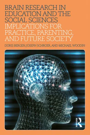
Brain Research in Education and the Social Sciences: Implications for Practice, Parenting, and Future Society provides practitioners, parents, and policy makers with research-based information and illustrative case studies about brain development across the lifespan. Neurotechnological advances that are contributing to a broader understanding of brain development and brain illnesses are discussed in a context specifically relevant to those working in education and the social sciences. The book enables readers to understand the societal implications of this expanding knowledge base and offers suggestions for future policies and practices that would make high-quality learning environments available to all students and individuals receiving care.
eBook - ePub
Brain Research in Education and the Social Sciences
Implications for Practice, Parenting, and Future Society
- 206 pages
- English
- ePUB (mobile friendly)
- Available on iOS & Android
eBook - ePub
Brain Research in Education and the Social Sciences
Implications for Practice, Parenting, and Future Society
About this book
Trusted by 375,005 students
Access to over 1.5 million titles for a fair monthly price.
Study more efficiently using our study tools.
Information
Topic
EducationSubtopic
Education General1
Understanding the Brain
Men ought to know that from the brain, and from the brain only, arise our pleasures, joys, laughter and jests, as well as our sorrows, pains, griefs and tears. Through it, in particular, we think, see, hear, and distinguish the ugly from the beautiful, the bad from the good, the pleasant from the unpleasant … it … makes us mad or delirious, inspires us with dread or fear, whether by night or by day, brings sleeplessness, inopportune mistakes, aimless anxieties, absent-mindedness, and acts that are contrary to habit.
—Hippocrates
The Sacred Disease, in Hippocrates, Trans. W. H. S. Jones (1923), Vol. 2, 175
The Sacred Disease, in Hippocrates, Trans. W. H. S. Jones (1923), Vol. 2, 175
A Brief History of the Study of the Brain
Recent emphasis on brain research suggests that study of the brain is a new phenomenon; however, structures and functions of the brain have been studied by physicians, philosophers, and researchers since earliest times. Archeologists have found what appears to be the deliberate drilling of holes in human skulls, perhaps to relieve pressure from brain injury. As early as 1700 BC, Egyptian doctors used surgery to treat brain injuries and observed connections between the central nervous system, sensation, and locomotion. In the mummification process, however, Egyptians preserved the heart and liver but not brain matter (Finger, 1994, 2001). Most information about the human brain was based upon study of dead animals and humans, so many assumptions about the human brain were erroneous. (e.g., human and sheep brains had similar cavities).
In 450 BC, Alcmaeon of Croton described the optic nerve as being a path of light to the brain (Gross, 1998). Early interest in the brain was evident in China, India, and Syria from 400 BC to 100 AD (Finger, 1994, 2001; Gross, 1998). The Chinese stated there was a connection between the eyes and the brain (i.e., the evil eye), and Nemesius of Syria proposed that basic sensory and motor functions were in the ventricles (cavities). This (incorrect) view was accepted throughout the middle ages. Greek and Roman philosophers and physicians also debated functions of the brain. Hippocrates drilled holes in the skulls of patients because he thought it would balance the humours of the brain and Galen identified the autonomic and sympathetic nervous systems, stating that wounds of the brain affect the mind. However, he concluded that the outer portion of the brain (the cortex) was just a covering with no functional importance (“cortex” means “rind” or “bark”). Although Plato saw the brain as the site of sensation and thought, Aristotle thought the heart was the major center of rationality and that the brain only cooled “humours” of the blood. This “cardiocentric” view prevailed throughout the middle ages. Aristotle stated that “spirit” circulated freely in the brain, via sensus communis, which came to be known as “common sense.”
Beginnings of Scientific Study
Prior to the thirteenth century, most physicians were associated with the Church, so doctors applied a mix of religious dogma and medicinal practices to brain injuries and diseases (Finger, 2001). In the mid-fourteenth century, most serious injuries (e.g., during wars) were not treated, as it was thought humours would be released into the brain, which, being untreatable, would kill the person. By the mid-1500s, medicine began to be based on anatomy and physiology, and physicians operated on severe head injuries with surgical tools and pharmacological means (Finger, 2001). Vesalius concluded that Galen’s earlier description of brain anatomy was erroneous and suggested revisions. Leonardo da Vinci’s illustrations of animal brains and his wax casts of brain ventricles offered precise anatomical information about brain structures and, although his information corrected earlier illustrations of the brain, it still did not challenge the incorrect idea that the ventricles were sources of sensory and motor functions.
The Renaissance opened a new world of brain study (Finger, 1994; Gross, 1998). Systematic study of relationships between brain and learning processes began in the seventeenth and eighteenth centuries. Using the microscope (an early technological advance), Malpighi studied the anatomy of the cortex, Willis suggested that the gyri (bulging folds of the cerebral cortex) controlled memory and will, and Descartes identified the pineal gland while still asserting that the brain and the mind were two different entities (Gross, 1998). By the 1700s, various areas of the brain had been identified as responsible for specific functions. For example, Legallois isolated the medulla as the respiratory center of the brain (Cheung, 2013), Swedenborg identified separate motor and sensory cortical areas (Fingers, 1994), Flourens reported that the cerebellum coordinated movement (Yildirim & Sarikcioglu, 2007), Broca showed that motor control of language was in the frontal cortex (Pierre-Paul Broca, 2000), and Wernicke identified the temporal lobe as the site for interpretation of spoken language (Finger, 1994). Unfortunately, Gall’s promotion of phrenology, an incorrect view asserting that skull shape and features were related to personality characteristics, gained prominence until it was attacked by Flourens, who stated that the brain acts as a whole to form intelligence. Although still discussed in some introductory psychology courses, phrenology was discounted by the late 1800s.
Prior to the 1900s, with the assistance of electrophysical recordings (another technological advance), Cajal established the “Neuron Doctrine,” stating that neurons were independent units composed of cell bodies, axons, and dendrites; Schwann identified the myelin sheaths (fat-like deposits) on neurons; and Sherrington found the “spaces” between axon and dendrites (the synapses) and studied how messages were transmitted across the synapses (Finger, 1994, 2001; Gross, 1998; Yuste, 2015). By the early 1900s, the theory that brain functions were specific to certain areas was challenged by the equipotentiality theory, which suggested that all areas of the brain contribute equally to behaviors (Lashley, 1950). Lashley later modified his theory to suggest that subareas of each region have some specific functions. Luria (1973) developed a competing idea of the brain as being composed of “functional systems,” which suggested that multiple areas of the brain function together to produce behaviors. Franz first investigated the brain’s relationship to learning by studying the brain’s ability to learn or relearn after it had been damaged (Finger, 1994, 2001). His findings suggested that new pathways could be developed after brain injury, supporting more integrated theories of brain functioning.
During the late twentieth century, major technological advances enabled brain researchers to engage in more precise study of the brain. The development of metabolic, electrophysiologic, magnetic, and neuropsychological methods for studying living brains has enabled researchers to learn much more about relationships between brain structures and functions (Zillmer, Spiers, & Culbertson, 2008). Information in this book is primarily derived from research using these newer procedures. They include metabolic procedures such as positron emission tomography (PET), single photon emission tomography (SPECT), and functional magnetic resonance imaging (fMRI); electrophysiological procedures such as EEG and ERP; and magnetic procedures such as magnetic source imaging (MSI), magnetic encephalography (MEG), and diffusion tensor imaging (DTI). Because the procedures are still being refined and extended, many of the implications of these findings for education and development are hypotheses only. The advantages and disadvantages of each procedure are shown in Table 1.1.
Metabolic Procedures
These procedures permit researchers to learn more about where thinking is occurring in the brain. They include positron emission tomography (PET), single-photon emission tomography (SPECT), and functional magnetic resonance imaging (fMRI). PET and SPECT procedures follow the flow and usage of radioactive compounds (e.g., oxygen or glucose for PET and technetium-99 or xenon-133 for SPECT) through the brain while the person is performing mental tasks. As the radioactive substance decays, a positron (positively charged electron-like particle) is emitted and tracked. High emissions from an area means high levels of oxygen or glucose are required in that area during cognitive activity; therefore, the areas of greatest activity during particular tasks (e.g., naming pictures, reading, memorizing) can be observed.
| Technique | Advantages | Disadvantages |
|---|---|---|
| PET/SPECT Positron emissions tomography/single-photon emission tomography | —Non-invasive —Able to localize brain activity —Able to use on young children | —Ethical questions of use of radioactive materials on the young —Range of isolation of activity is centimeter range (not precise) —Brief time span before decay of signal —Expensive |
| fMRI Functional magnetic resonance imaging | —Noninvasive —No exposure to radiation —Precise; isolates areas in range of millimeters —Fast | —Participants must be very still —High level of noise may be painful —Cramped environment —Expensive |
| EEG Electroencephalogram | —Noninvasive —Sensitive to state changes —Relatively inexpensive | —Gives general information; not really suited to study of cognitive processes —Poor spatial and temporal resolution |
| ERP Event-related potentials | —Can examine cognitive activity —Each signal marked by specific event/stimulus, so more accurate temporal information —More electrodes, so more accurate spatial information —Many measures in brief time | —Errors occur if extraneous movement (muscles, eyes), resulting in loss of trial or cases |
| MSI/MEG Magnetic source imaging/ encephalography | —Fast enough to track neural signals —Tracks neural pathways directly —Yields good spatial (in millimeters) and temporal (in milliseconds) data | —Only reads signals near brain surface —Extremely sensitive to outside magnetic fields (moving metal objects) —Expensive |
| DTI Diffusion tensor imaging | —Noninvasive —More sensitive to white matter injury of the brain than other imaging techniques —Highly sensitive to tears in white matter or diffuse axonal injury (DAI) —Can be used along with neuropsychological testing to show evidence of cognitive decline —Can help predict recovery times for concussion patients | —Similar to other MRI tests —Expensive —Highly sensitive to distortion based on movement by the patient —Images can be blurry due to its low spatial resolution |
| Neuropsychological assessment | —Noninvasive —Tasks can be used in animal research to make inferences abo... |
Table of contents
- Cover
- Title
- Copyright
- Dedication
- Contents
- List of Figures
- List of Tables
- Introduction
- 1 Understanding the Brain
- 2 The Brain’s Functions and Structures
- 3 Prenatal Brain Development as a Foundation for Learning
- 4 Brain Development and Learning in the Infant and Toddler Years
- 5 Brain Development and Learning in the Preschool Years
- 6 Brain Development and Learning in the Elementary Years
- 7 Brain Development and Learning in the Middle Childhood Years
- 8 Brain Development and Learning in the Adolescent Years
- 9 Brain Development and Learning in the Adult Years
- 10 Using Neuroimaging and Neurodiagnostic Techniques to Understand and Improve Brain Functioning
- 11 Using EEG and ERP Methodology to Understand and Improve Brain Functioning
- 12 Influence of Brain Research on Clinical and Educational Practice
- 13 Evaluating Professional Practices From a Brain Research Perspective
- 14 Futurist Issues and Predictions for Human Brain Development
- Glossary of Brain and Nervous System Terms
- Index
Frequently asked questions
Yes, you can cancel anytime from the Subscription tab in your account settings on the Perlego website. Your subscription will stay active until the end of your current billing period. Learn how to cancel your subscription
No, books cannot be downloaded as external files, such as PDFs, for use outside of Perlego. However, you can download books within the Perlego app for offline reading on mobile or tablet. Learn how to download books offline
Perlego offers two plans: Essential and Complete
- Essential is ideal for learners and professionals who enjoy exploring a wide range of subjects. Access the Essential Library with 800,000+ trusted titles and best-sellers across business, personal growth, and the humanities. Includes unlimited reading time and Standard Read Aloud voice.
- Complete: Perfect for advanced learners and researchers needing full, unrestricted access. Unlock 1.5M+ books across hundreds of subjects, including academic and specialized titles. The Complete Plan also includes advanced features like Premium Read Aloud and Research Assistant.
We are an online textbook subscription service, where you can get access to an entire online library for less than the price of a single book per month. With over 1.5 million books across 990+ topics, we’ve got you covered! Learn about our mission
Look out for the read-aloud symbol on your next book to see if you can listen to it. The read-aloud tool reads text aloud for you, highlighting the text as it is being read. You can pause it, speed it up and slow it down. Learn more about Read Aloud
Yes! You can use the Perlego app on both iOS and Android devices to read anytime, anywhere — even offline. Perfect for commutes or when you’re on the go.
Please note we cannot support devices running on iOS 13 and Android 7 or earlier. Learn more about using the app
Please note we cannot support devices running on iOS 13 and Android 7 or earlier. Learn more about using the app
Yes, you can access Brain Research in Education and the Social Sciences by Doris Bergen,Joseph Schroer,Michael Woodin in PDF and/or ePUB format, as well as other popular books in Education & Education General. We have over 1.5 million books available in our catalogue for you to explore.