The brain controls every aspect of human life. It is responsible for everything we do and think, from basic bodily functions, such as breathing, blood pressure and sleep control, to the expression of personality and emotions, and control of thoughts and behaviour. It controls our ability to move, sense things, plan and organise, and communicate, etc. It may therefore seem surprising that few of us understand or have been taught much about the brain, as the all-important part of our body that enables us to know who we are and what we do.
It is helpful to recognise what the brain does and how it works in order to appreciate what might have happened when it is injured and therefore may not work efficiently.
The following description of the basic structure and function of the brain is presented in a simplistic, non-technical way. This is not meant to trivialise the topic, but to provide an accessible and introductory guide to readers who may not be familiar with this. More detailed information about brain development and functioning can be found in Reed and Warner-Rogers (2008).
Structure of the brain
The central nervous system is made up of the brain and the spinal cord. The spinal cord is a bundle of nerves that connects the brain to other parts of the body. It receives signals from all parts of the body, relays information upwards and downwards from the brain and is protected by a series of bones called vertebrae, collectively known as the vertebral column. Although the spinal cord is susceptible to damage, the results of a spinal cord injury are very different from those of injuries to the brain and are therefore not referred to here.
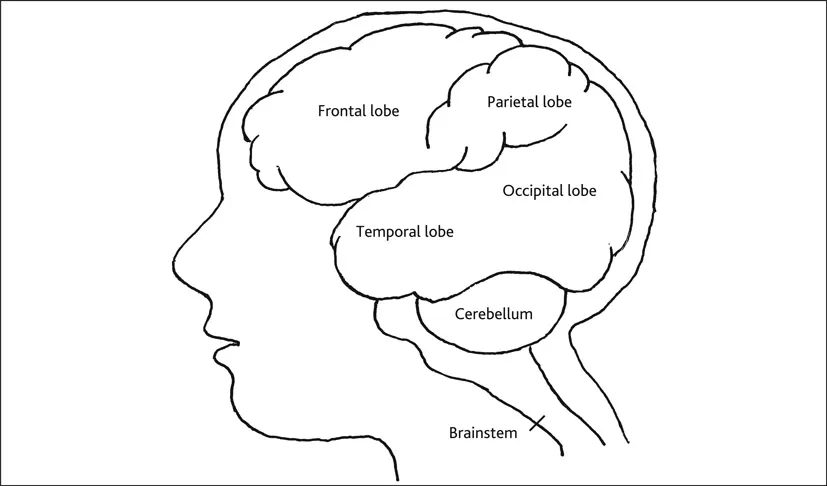
Figure 1.1 Structure of the brain
The brain is a complex structure. The terminology that has evolved to describe it is also complex and most structures have acquired several alternative labels derived from Greek, Latin, English or French.
The brain can be described as having three major parts: the brainstem, the cerebellum and the cerebral cortex. However, although each area has specific functions, they are not distinct or separate working units but an organisation of multiple and interconnected systems that are heavily dependent on each other (see Figure 1.1).
The brainstem is the lowest part of the brain, which tapers into the spinal cord. In lay terms it can be said to function like a telecommunications cable (Powell 1994): a collection of nerves and fibres carrying all messages backwards and forwards between the brain and the rest of the body. However, the brainstem is also responsible for basic bodily functions essential to life such as breathing, heart rate and blood pressure, and plays a vital role in basic attention, arousal and consciousness.
The cerebellum – meaning ‘little brain’ – is a cauliflower-like structure that sits under the cerebral cortex. It controls and co-ordinates bodily movement and muscle tone – the degree of muscle tension during rest or in response to stretching. It develops and stores information regarding the motor – i.e. physical – skills that enable us to walk, run, climb, ride bikes, carry out fine motor activities such as handwriting or using a knife and fork, and many other physical activities. The cerebellum helps control direction, rate, force and smoothness of movement. It is situated just above the brainstem at the back of the brain and is thus relatively well protected from traumatic injury compared with other parts of the brain.
The cerebral cortex is the largest part of the brain, dedicated to the highest levels of thinking, moving and acting, and makes up seven-tenths of the entire nervous system in humans. It is shaped like a large walnut: its surface is convoluted with many deep furrows – sulci – and raised surfaces – gyri – enabling increased surface area within the confines of a relatively small skull. Neuroscientists believe that it is the cerebral cortex that sets us apart from all other creatures. The outer layer of the cortex has a greyish appearance and is often referred to as ‘grey matter’.
The cerebral cortex is divided into two halves: the right and left hemispheres. Although similar in appearance, they have different functions. The left cerebral hemisphere controls the right side of the body and is usually responsible for speech and language functions; the right cerebral hemisphere controls the left side of the body and is usually responsible for processing visual and spatial information and some other non-verbal skills.
The differences in specialities in the right and left hemispheres can indicate that an injury to the left side of the brain would be more likely to result in language difficulties and/or right-sided problems, while injury to the right side of the brain may produce difficulties with visual perception and/or left-sided problems. However, it is important to note that, although some brain injuries may only affect a specific area or areas, many lead to more widely spread damage (see Chapter 2).
The two hemispheres are linked by nerve fibres, collectively called the corpus callosum, which serve as a bridge or channel of communication between the hemispheres.
Each hemisphere is further divided into four parts, or lobes: occipital, parietal, temporal and frontal. These are named after the overlying bones of the skull. Each lobe is associated with different aspects of functioning:
- Occipital lobes, at the very back of the head, are involved in processing visual information. This includes visual acuity – often just termed ‘vision’ – and the way visual information is interpreted, e.g. colour, word or object recognition.
- Parietal lobes are located at the back and top of the head, behind the frontal lobes and above the temporal lobes. They are responsible for the processing of information about body sensation – touch, pressure, temperature and pain – as well as the integration of visual and auditory information and an understanding of spatial relationships.
- Temporal lobes are located under the temples and are important for hearing and many aspects of memory. They are crucially involved in certain processes relating to attention and language, to musical ability, and to facial and other aspects of visual recognition. An area of the brain called Wernicke’s Area spans the temporal and parietal lobes and is partly responsible for our understanding and production of language. Deep within each temporal lobe is also a structure called the hippocampus – named thus as its shape is said to resemble a seahorse. This plays an important role in memory and is linked with other areas involved with emotion.
- Frontal lobes are located behind the forehead. They are relatively immature during childhood and develop over an extended period into early adulthood. They are extremely vulnerable to injury because of their location at the front of the head (McAllister 2011). The frontal lobes are responsible for some motor functions and some aspects of memory but are particularly important in respect of ability in problem solving, initiation, judgement, impulse control, etc. They act as ‘behavioural regulators’, planning and evaluating behavioural responses. These, along with other behaviours, are known as ‘executive functions’ (see Chapter 4). The frontal lobes also contain a structure known as Broca’s Area that is associated with our ability to speak and also, to some extent, to understand language (Caplan 2006).
There are other structures deep within the centre of the brain that have important functions, such as those relating to emotion and sleep control.
Brain development
Growth before birth
Initially, the human embryo consists of only a few primitive cells which develop into the body and all its vital organs. Brain tissue is made up primarily of nerve cells – or neurons – and supporting cells called glial – meaning glue – cells. During a very early stage of foetal development – week three of gestation – something called the neural tube is formed, which is mainly comprised of neural stem cells and is filled with cerebrospinal fluid. Neurons – and glial cells – are produced; this is called neurogenesis. These initially form a basic front, mid- and hindbrain and then the newly formed neurons move to form other brain structures. This migration is guided partly by paths created by glial cells, which direct the neurons to their ultimate location. When they reach their destination, the cells start to specialise.
By week nine of foetal development, the brain has already taken on its adult shape, with the typical convolutions – folds – of the cortex. The growth and development of the brain in the womb is more accelerated than any other part of the foetus; a newborn baby’s brain is one-third of the adult brain weight, although the baby’s overall weight is one-twentieth as heavy as the adult it will become – the average weight of a brain in a newborn baby is 450 g and by adulthood the average weight is 1,400 g. As a result, when babies are born their heads are very large in relation to the rest of their body.
Neurogenesis continues during the first few months of life. It was previously thought that after this initial production of neural cells during early development, no new neurons could be generated, but we now know that limited natural neurogenesis continues in adults, in certain brain regions (Paspala et al. 2011). Currently this has been identified primarily in the hippocampus – see description of temporal lobes, pages 6–7. There is also considerable current interest and research regarding the potential use of stem cells in cell replacement therapy.
Prenatal neural development is thought to be mainly genetically determined. Interruptions to development during this period, such as trauma or infection, are likely to have a significant impact on cerebral structure.
Growth after birth
A newborn baby’s brain has about a hundred billion cells, but they have only just begun to develop the connections that enable the brain to be an organised and integrated system. The nerve cells, or neurons, continue to grow, sprouting thin fibres, called dendrites, like wide, ...